Polyclonal Antibodies
Anti-IRAK4 Polyclonal Antibody for WB, IF/ICC, ELISA - Q9NWZ3
Item Number : CM0024586
Price varies based on specs and customizations
- Application
- WB, IF/ICC, ELISA
- Cross Reactivity
- Human, Mouse, Rat
- Protein Weight
- 52kDa

Shipping:
Contact us to get shipping details Enjoy On-time Dispatch Guarantee.
Why Choose Us
Easy ordering process, quality products, and dedicated support for your business success.
Core Product Specifications and Parameters
| Parameter | Value |
|---|---|
| Product Name | IRAK4 Rabbit pAb |
| Remarks/Alias | IPD1; IMD67; REN64; IRAK-4; NY-REN-64; IRAK4 |
| Species | Human |
| GeneID (human) | 51135 |
| Immunogen | Recombinant protein |
| Source | Rabbit |
| Category | Polyclonal Antibodies |
| Application | WB, IF/ICC, ELISA |
| Cross Reactivity | Human, Mouse, Rat |
| SWISS | Q9NWZ3 |
| Protein Weight | 52kDa |
| Shipping | Ice bag |
Biological Background: IRAK4 Function and Localization
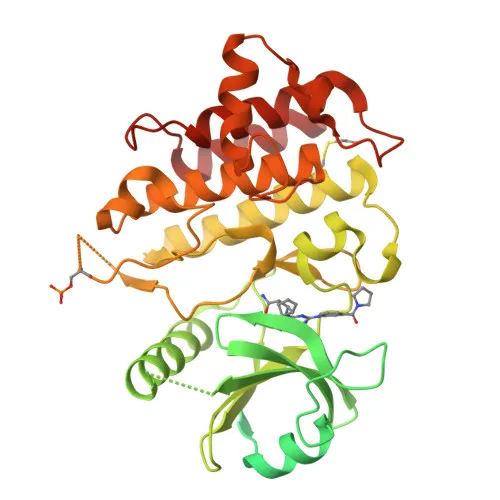
- IRAK4 (Interleukin-1 receptor-associated kinase 4) is a serine/threonine kinase essential for innate immune signaling, encoded by the IRAK4 gene in humans. It is also known as renal carcinoma antigen NY-REN-64.
- Upon activation of Toll-like receptors (TLRs) or interleukin-1 receptor (IL-1R), IRAK4 is recruited by MYD88 to form the Myddosome complex with IRAK2, initiating downstream signal transduction. Related references: PMID:17878374
- IRAK4 phosphorylates IRAK1, which then undergoes autophosphorylation and forms a complex with TRAF6 and TAK1; it also phosphorylates Pellino proteins (PELI1, PELI2, PELI3) to promote polyubiquitination of IRAK1, ultimately leading to NF-κB nuclear translocation and activation of inflammatory gene expression.
- Alternatively, IRAK4 phosphorylates TIRAP to promote its ubiquitination and degradation, and phosphorylates NCF1 to regulate NADPH oxidase activation after LPS stimulation, suggesting a role in microbicidal responses.
- Subcellularly, IRAK4 localizes primarily to the cytoplasm, consistent with its function in signalosome assembly at membrane-proximal sites.
- Keywords associated with IRAK4 include kinase, nucleotide-binding, magnesium, acetylation, alternative splicing, and disease variants; mutations in IRAK4 are linked to primary immunodeficiency disorders such as IPD1 and IMD67, characterized by increased susceptibility to pyogenic bacterial infections.
Experimental Guidance and Technical Tips
- The antibody was raised against an N-terminal fragment (aa 1-200) covering the death domain of human IRAK4; it may recognize both full-length and N-terminally truncated isoforms if present.
- For Western blotting, expect a band at approximately 52 kDa in lysates from human, mouse, or rat cell lines; molecular weight can vary slightly due to phosphorylation or gel conditions.
- In immunofluorescence/ICC, cytoplasmic localization should be observed; use appropriate fixation and permeabilization protocols.
- For ELISA, consider antibody titration and optimize coating antigen concentration to achieve the best signal-to-noise ratio.
- Cross-reactivity with mouse and rat IRAK4 is indicated by the supplier; validation in the specific sample systems is recommended before large-scale studies.
- To study IRAK4 activation, stimulate cells with IL-1β or TLR ligands (e.g., LPS) and assess phosphorylation status or complex formation using co-immunoprecipitation.
CamelBio: Your One-Stop Sourcing Bridge
CamelBio provides diagnostic manufacturers and research labs with reliable access to high-quality IVD raw materials such as this anti-IRAK4 polyclonal antibody, which is suitable for developing immunoassays targeting innate immune pathways. Our comprehensive portfolio includes validated antibody pairs, optimized monoclonal and polyclonal antibodies, and specialized ancillary reagents, streamlining your workflow from research to clinical application. Contact us to discuss custom sourcing solutions for rare targets or bulk reagent needs.
REQUEST A QUOTE
Our professional team will reply to you within one business day. Please feel free to contact us!