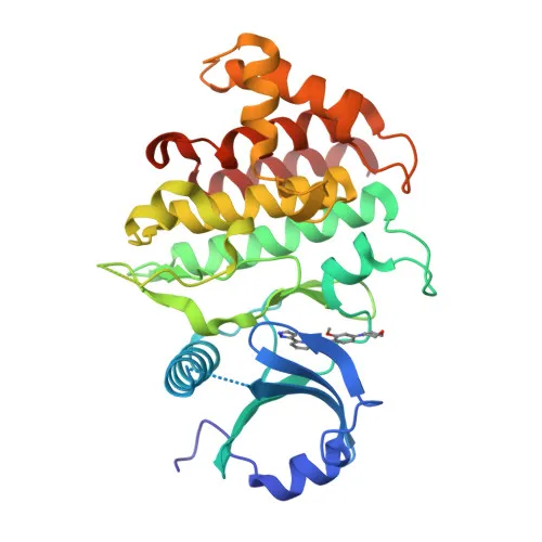
Polyclonal Antibodies
Anti-IRAK1 Polyclonal Antibody for WB, IF/ICC, ELISA - P51617
Item Number : CM0017520
Price varies based on specs and customizations
- Application
- WB, IF/ICC, ELISA
- Cross Reactivity
- Human, Mouse, Rat
- Protein Weight
- 77kDa

Shipping:
Contact us to get shipping details Enjoy On-time Dispatch Guarantee.
Why Choose Us
Easy ordering process, quality products, and dedicated support for your business success.
Core Product Specifications and Parameters
| Parameter | Value |
|---|---|
| Product Name | IRAK1 Rabbit pAb |
| Remarks/Alias | IRAK; pelle; IRAK1 |
| Species | Human |
| GeneID (human) | 3654 |
| GeneID | 3654 |
| Immunogen | Recombinant fusion protein containing a sequence corresponding to amino acids 424-633 of human IRAK1 (NP_001020414.1) |
| Source | Rabbit |
| Category | Polyclonal Antibodies |
| Application | WB, IF/ICC, ELISA |
| Cross Reactivity | Human, Mouse, Rat |
| SWISS | P51617 |
| Protein Weight | 77kDa |
| Shipping | Ice bag |
Biological Background: IRAK1 Function and Localization
- IRAK1 (interleukin-1 receptor-associated kinase 1) is a serine/threonine kinase essential for initiating innate immune responses, acting as a key mediator in Toll-like receptor (TLR) and IL-1 receptor signaling pathways.
- Upon TLR/IL-1R activation, IRAK1 is rapidly recruited by MYD88, phosphorylated by IRAK4, and undergoes autophosphorylation, which triggers downstream signaling events.
- IRAK1 phosphorylates Pellino E3 ubiquitin ligases (PELI1, PELI2, PELI3), promoting polyubiquitination of IRAK1 itself, leading to recruitment of the IKK complex and subsequent NF-kB nuclear translocation.
- It also phosphorylates TIRAP to promote its ubiquitination and degradation, and directly phosphorylates IRF7 to drive type I interferon production, establishing an antiviral state.
- Subcellular localization includes cytoplasm, nucleus, and lipid droplets; sumoylation of IRAK1 facilitates nuclear translocation where it phosphorylates STAT3.
- Two main isoforms exist: isoform 1 is more strongly expressed than isoform 2, both ubiquitously present in human tissues.
- IRAK1 is involved in host-virus interactions and undergoes various post-translational modifications including phosphorylation, ubiquitination, and sumoylation.
Experimental Guidance and Technical Tips
- The immunogen corresponds to amino acids 424-633 of human IRAK1, a region conserved across human, mouse, and rat, supporting the observed cross-reactivity.
- For Western blot, consider using whole cell lysates from TLR/IL-1R-stimulated immune cells (e.g., THP-1, RAW264.7) to detect activation-dependent bands near 77 kDa; basal expression may vary.
- In IF/ICC experiments, optimize fixation and permeabilization conditions; endogenous IRAK1 may localize to both cytoplasm and nucleus depending on cell type and stimulation.
- For ELISA, pairing with a matched detection antibody (e.g., against a different epitope) may improve sensitivity; validate in the relevant sample matrix.
- As a polyclonal antibody, lot-to-lot variability is possible; include positive (e.g., stimulated cells) and negative controls in each assay.
CamelBio: Your One-Stop Sourcing Bridge
CamelBio streamlines your IVD raw material sourcing by offering this high-quality anti-IRAK1 polyclonal antibody alongside validated antibody pairs, optimized monoclonal antibodies, and bulk ancillary reagents. Our one-stop platform supports manufacturers and researchers in developing robust immunoassays and diagnostic kits for innate immunity and inflammatory disease profiling. Let CamelBio be your reliable partner in accelerating from concept to clinic.
REQUEST A QUOTE
Our professional team will reply to you within one business day. Please feel free to contact us!