Polyclonal Antibodies
Anti-IFNLR1 Rabbit Polyclonal Antibody for WB, ELISA - Q8IU57
Item Number : CM0016960
Price varies based on specs and customizations
- Application
- WB, ELISA
- Cross Reactivity
- Human, Mouse, Rat
- Protein Weight
- 58kDa

Shipping:
Contact us to get shipping details Enjoy On-time Dispatch Guarantee.
Why Choose Us
Easy ordering process, quality products, and dedicated support for your business success.
Core Product Specifications and Parameters
| Parameter | Value |
|---|---|
| Product Name | IFNLR1 Rabbit pAb |
| Remarks/Alias | IFNLR; LICR2; IL28RA; CRF2/12; IL-28R1; IFNLR1 |
| Species | Human |
| GeneID (Human) | 163702 |
| GeneID | 163702 |
| Immunogen | Recombinant protein / Recombinant fusion protein containing a sequence corresponding to amino acids 21-228 of human IFNLR1 (NP_734464.1). |
| Source | Rabbit |
| Category | Polyclonal Antibodies |
| Application | WB, ELISA |
| Cross Reactivity | Human, Mouse, Rat |
| SWISS | Q8IU57 |
| Protein Weight | 58kDa |
| Shipping | Ice bag |
Biological Background: IFNLR1 Function and Localization
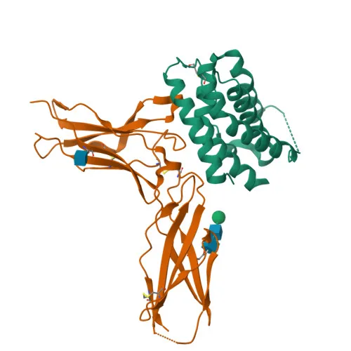
- IFNLR1 (Interferon lambda receptor 1), also known as IL28RA, CRF2-12, or LICR2, is the ligand-binding subunit of the functional receptor complex for type III interferons (IFN-λ), which includes IFNL2 and IFNL3.
- It dimerizes with IL10RB to form a functional receptor that mediates antiviral activity by activating the JAK/STAT signaling pathway, leading to the expression of interferon-stimulated genes (ISGs).
- The receptor determines cell type specificity of lambda interferon action, being predominant in epithelial cells of the respiratory, gastrointestinal, and reproductive tracts, thereby limiting responses primarily to these tissues.
- Subcellular localization is at the plasma membrane as a single-pass membrane protein, consistent with its role in extracellular ligand binding and signal transduction.
- The gene undergoes alternative splicing, producing isoforms with potential functional differences.
- Post-translational modifications include disulfide bond formation, glycosylation, and ubiquitination (Ubl conjugation), which may regulate receptor stability and signaling.
- Tissue expression is widespread, but functional impact is most prominent in barrier epithelia where it plays a significant role in antiviral immune defense, particularly in the intestinal epithelium.
- Structurally, IFNLR1 belongs to the class II cytokine receptor family, featuring a signal peptide and a transmembrane helix.
Experimental Guidance and Technical Tips
- For Western blot, the predicted molecular weight is 58kDa. Consider using a reducing SDS-PAGE and standard PVDF membrane transfer. Validate with appropriate positive control lysates from human, mouse, or rat tissues rich in epithelial cells (e.g., intestinal epithelium).
- For ELISA, this polyclonal antibody can be used as a detection or capture reagent. Ensure to titrate the antibody to optimize signal-to-noise ratio. Pair with a validated anti-IFNLR1 monoclonal antibody if a sandwich ELISA is intended.
- The immunogen encompasses a large extracellular fragment, so the antibody may recognize both full-length and soluble splice variants if present. Consider testing in samples with known IFNLR1 expression to assess specificity.
- Since cross-reactivity is reported for human, mouse, and rat, verify reactivity in target species by sequence alignment of the immunogen region. Minor differences may affect sensitivity.
CamelBio: Your One-Stop Sourcing Bridge
CamelBio offers a comprehensive selection of IVD raw materials, including validated antibodies, antigens, and ancillary reagents to support your immunoassay development. For researchers focusing on antiviral defense and cytokine signaling, this anti-IFNLR1 polyclonal antibody is part of our extensive portfolio of immune-related reagents. Contact us for customized bulk orders and technical consultation to accelerate your diagnostic projects.
REQUEST A QUOTE
Our professional team will reply to you within one business day. Please feel free to contact us!