Polyclonal Antibodies
Anti-ICAM-1/CD54 Rabbit Polyclonal Antibody for WB, IHC-P, IF/ICC, ELISA - P05362
Item Number : CM0027566
Price varies based on specs and customizations
- Application
- WB, IHC-P, IF/ICC, ELISA
- Cross Reactivity
- Human, Mouse, Rat
- Protein Weight
- 58kDa

Shipping:
Contact us to get shipping details Enjoy On-time Dispatch Guarantee.
Why Choose Us
Easy ordering process, quality products, and dedicated support for your business success.
Core Product Specifications and Parameters
| Parameter | Value |
|---|---|
| Product Name | ICAM-1/CD54 Rabbit pAb |
| Remarks/Alias | BB2; CD54; P3.58; ICAM-1/CD54 |
| Species | Human |
| GeneID (Human) | 3383 |
| GeneID | 3383 |
| Immunogen | Recombinant fusion protein containing a sequence corresponding to amino acids 28-480 of human ICAM-1/CD54 (NP_000192.2) |
| Source | Rabbit |
| Category | Polyclonal Antibodies |
| Application | WB, IHC-P, IF/ICC, ELISA |
| Cross Reactivity | Human, Mouse, Rat |
| SWISS | P05362 |
| Protein Weight | 58kDa |
| Shipping | Ice bag |
Biological Background: ICAM-1/CD54 Function and Localization
- ICAM-1 is a cell adhesion molecule that serves as a ligand for the integrin LFA-1 (ITGAL:ITGB2), mediating leukocyte adhesion and providing a calibrated system for T-cell killing. Related references: PMID:3086451 PMID:3340213 PMID:38195629
- It also functions as a receptor for MAC-1 (ITGAM:ITGB2), enabling adhesion between stimulated neutrophils and endothelial cells. Related references: PMID:1980124
- During leukocyte trans-endothelial migration, ICAM1 engagement triggers endothelial apical cup assembly via ARHGEF26/SGEF and RHOG. Related references: PMID:17875742
- In epithelial cells, ICAM-1 promotes cell aggregation through interaction with MUC1. Related references: PMID:11173916
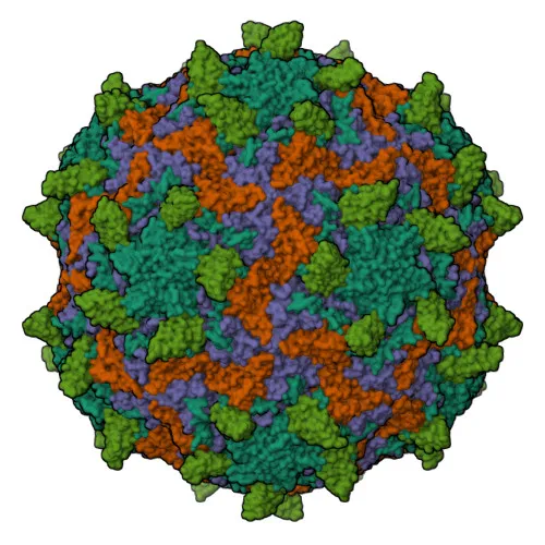
- (Microbial infection) ICAM-1 acts as a receptor for major group rhinoviruses (A, B) and Coxsackievirus A21, facilitating viral entry.
- Upon HHV-8 infection, ICAM-1 is degraded by viral E3 ubiquitin ligase MIR2 to evade immune clearance.
- ICAM-1 is a single-pass type I membrane protein localized at the cell membrane; it is expressed on vascular endothelial cells, thymic epithelial cells, fibroblasts, and certain leukocytes, with low basal expression on peripheral blood leukocytes. Related references: PMID:3086451
- Post-translational modifications include disulfide bond formation, N-linked glycosylation, phosphorylation, and ubiquitination.
Experimental Guidance and Technical Tips
- The immunogen spans the extracellular domain (aa 28-480), suggesting that this polyclonal antibody may recognize surface-exposed epitopes; consider its potential for flow cytometry or live-cell staining with appropriate validation.
- For Western blotting, expect a band near the predicted molecular weight of ~58 kDa; however, glycosylation often results in a higher apparent molecular weight (75–100 kDa). Optimize reducing conditions and loading controls.
- For immunohistochemistry (paraffin sections), heat-induced antigen retrieval may improve detection of this membrane protein.
- In immunofluorescence/ICC, include a permeabilization step for intracellular detection or omit it to specifically visualize surface localization.
- The antibody cross-reacts with mouse and rat; validate performance in these species using relevant positive control tissues (e.g., inflamed endothelium) and appropriate negative controls.
CamelBio: Your One-Stop Sourcing Bridge
CamelBio provides diagnostic manufacturers, laboratories, and research institutes with comprehensive access to IVD raw materials, technical services, and consulting—from concept to clinic. For ICAM-1/CD54 research and assay development, we offer validated antibody pairs, recombinant proteins, and optimized polyclonal antibodies like this one, alongside bulk ancillary reagents and custom sourcing for rare targets.
Product Datasheet
Anti-ICAM-1/CD54 Rabbit Polyclonal Antibody for WB, IHC-P, IF/ICC, ELISA - P05362
REQUEST A QUOTE
Our professional team will reply to you within one business day. Please feel free to contact us!