Monoclonal Antibodies
Anti-Human IL-17RB Monoclonal Antibody () for Flow Cytometry - Q9NRM6
Item Number : CM0012655
Price varies based on specs and customizations
- Application
- FC
- Cross Reactivity
- Human
- Protein Weight
- 31kDa/55kDa

Shipping:
Contact us to get shipping details Enjoy On-time Dispatch Guarantee.
Why Choose Us
Easy ordering process, quality products, and dedicated support for your business success.
Core Product Specifications and Parameters
| Parameter | Value |
|---|---|
| Product Name | Rabbit anti-Human IL-17RB mAb |
| Remarks/Alias | IL17RB; CRL4; EVI27; IL17BR; IL17RH1; interleukin-17 receptor B |
| Species | Human |
| Gene ID | 55540 |
| Immunogen | Recombinant fusion protein containing amino acids 18-289 of human IL-17RB (NP_061195.2) |
| Source | Rabbit |
| Category | Monoclonal Antibodies |
| Application | FC |
| Cross Reactivity | Human |
| SWISS | Q9NRM6 |
| Protein Weight | 31kDa/55kDa |
| Shipping | Ice bag |
Biological Background: IL-17RB Function and Localization
- IL-17RB (Interleukin-17 receptor B), also known as IL-17Rh1, CRL4, or EVI27, is a single-pass transmembrane receptor encoded by the IL17RB gene on human chromosome 3.
- It functions as a receptor for the pro-inflammatory cytokines IL-17B and IL-17E (IL-25), mediating downstream signaling that may regulate hematopoietic cell growth and differentiation.
- The receptor contains a signal peptide, an extracellular domain, a transmembrane helix, and a cytoplasmic tail; it undergoes alternative splicing to generate secreted isoforms alongside the membrane-bound form.
- IL-17RB is localized primarily at the cell membrane, but a soluble, secreted form can also be generated by alternative splicing.
- Tissue expression is prominent in several endocrine tissues, including fetal and adult liver, kidney, pancreas, testis, colon, brain, and small intestine, but it is not detected in peripheral blood leukocytes or lymphoid organs.
- Post-translational modifications include N-linked glycosylation, as annotated in UniProt, consistent with its passage through the secretory pathway.
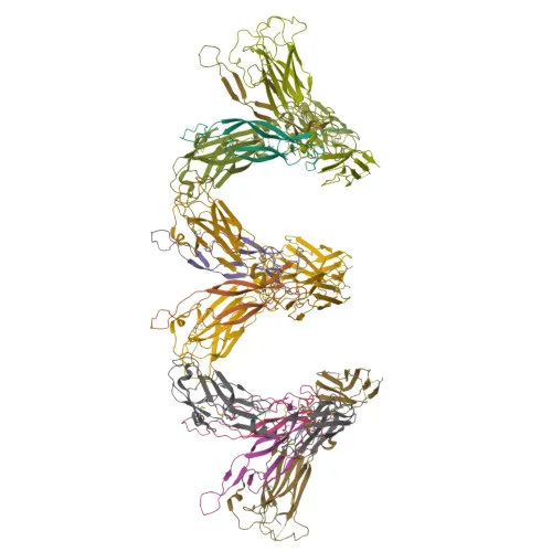
- Structurally, the receptor has been resolved by X‑ray crystallography, revealing features critical for ligand binding and specificity.
Experimental Guidance and Technical Tips
- This -conjugated antibody was raised against a recombinant protein spanning the extracellular domain (amino acids 18–289), a region involved in ligand recognition. For flow cytometry, consider titrating the antibody to establish an optimal staining concentration using IL-17RB‑positive cells (e.g., certain endocrine or epithelial lines) and a negative control.
- Since IL-17RB exhibits alternative splicing and potential secretion, ensure that membrane-bound expression is confirmed in your target sample system; stimulation with IL-17B/E or pro‑inflammatory conditions may upregulate surface expression.
- The antibody's reactivity is validated in human species; cross-reactivity with other species is not expected based on immunogen design, but users should verify for non-human applications.
- For multicolor flow panels, note that excites at ~650 nm and emits in the far‑red range; compensate appropriately and include Fc receptor blocking if using primary cells.
- Protein weight markers of 31 kDa and 55 kDa likely correspond to unglycosylated and glycosylated forms, respectively; western blot confirmation under reducing conditions may reveal similar bands if other detection formats are pursued.
CamelBio: Your One-Stop Sourcing Bridge
CamelBio provides diagnostic manufacturers, CROs, and research laboratories with integrated access to a broad portfolio of IVD raw materials. For studies targeting interleukin receptors like IL-17RB, we offer monoclonal antibodies, validated antibody pairs, and ancillary reagents—all from a single, quality-driven supply chain. Our team supports rare target sourcing and bulk procurement, helping streamline your transition from assay development to clinical application.
Product Datasheet
Anti-Human IL-17RB Monoclonal Antibody () for Flow Cytometry - Q9NRM6
REQUEST A QUOTE
Our professional team will reply to you within one business day. Please feel free to contact us!