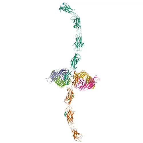
Monoclonal Antibodies
Anti-Human E-Cadherin/CD324 Monoclonal Antibody for Flow Cytometry - P12830
Item Number : CM0011849
Price varies based on specs and customizations
- Application
- FC
- Cross Reactivity
- Human
- Protein Weight
- 90kDa/97kDa

Shipping:
Contact us to get shipping details Enjoy On-time Dispatch Guarantee.
Why Choose Us
Easy ordering process, quality products, and dedicated support for your business success.
Core Product Specifications and Parameters
| Parameter | Value |
|---|---|
| Product Name | Rabbit anti-Human E-Cadherin/CD324 mAb |
| Remarks/Alias | UVO; CDHE; ECAD; LCAM; Arc-1; BCDS1; CD324 |
| Species | Human |
| GeneID (human) | 999 |
| GeneID | 999 |
| Immunogen | Recombinant fusion protein containing a sequence corresponding to amino acids 155-707 of human E-Cadherin/CD324 (NP_004351.1) |
| Source | Rabbit |
| Category | Monoclonal Antibodies |
| Application | FC |
| Cross Reactivity | Human |
| SWISS | P12830 |
| Protein Weight | 90kDa/97kDa |
| Shipping | Ice bag |
Biological Background: E-Cadherin Function and Localization
- Encoded by the CDH1 gene, cadherin-1 (E-cadherin) is a calcium-dependent cell adhesion protein that mediates homophilic cell-cell interactions, contributing to the sorting of heterogeneous cell types and the maintenance of epithelial integrity. Related references: PMID:11976333
- It organizes radial actin fiber structures and modulates cellular responses to contractile forces via its interaction with AMOTL2, thereby influencing epithelial mobility and proliferation.
- E-cadherin facilitates the early stages of desmosome assembly by recruiting DSG2 and DSP to cell-cell junctions, coupling adherens junctions with desmosomal complexes. Related references: PMID:29999492
- The protein is predominantly localized at the cell membrane, particularly at adherens junctions and desmosomes, but can also be found in the cytoplasm, endosomes, and the trans-Golgi network. A secreted form has been identified as well.
- Alternative splicing, proteolytic cleavage, and post-translational modifications such as phosphorylation and ubiquitination generate multiple isoforms and functional fragments with distinct roles in signaling and degradation.
- E-cadherin acts as a potent invasion suppressor in epithelial cells and serves as a ligand for integrin αE/β7 on intraepithelial lymphocytes.
- In amyloid metabolism, the C-terminal fragment (E-Cad/CTF2) promotes non-amyloidogenic degradation of APP, reducing the production of neurotoxic Aβ peptides.
- Pathologically, E-cadherin is hijacked as a receptor for Listeria monocytogenes internalin A, promoting bacterial uptake into host cells.
Experimental Guidance and Technical Tips
- The immunogen spans the extracellular domain (amino acids 155–707), ensuring recognition of surface-exposed epitopes suitable for live-cell flow cytometry applications.
- For flow cytometry (FC), titrate the antibody to determine the optimal staining concentration in your specific cell system. Include appropriate isotype controls and confirm specificity using E-cadherin knockout or knockdown models if available.
- Consider that E-cadherin is readily cleaved or shed; surface expression can be influenced by culture conditions, cell density, and fixation/permeabilization protocols. Validate detection parameters under conditions relevant to your experimental model.
- Because E-cadherin forms calcium-dependent homodimers, ensure staining buffers contain physiological calcium concentrations to preserve native conformation and epitope accessibility.
CamelBio: Your One-Stop Sourcing Bridge
CamelBio offers comprehensive access to IVD raw materials, including optimized monoclonal antibodies, bulk ancillary reagents, and custom sourcing for rare targets. This E-cadherin product supports researchers exploring epithelial cell biology, cancer metastasis, and cell adhesion. From concept to clinic, CamelBio serves as your trustworthy procurement partner for high-quality immunoreagents.
Product Datasheet
Anti-Human E-Cadherin/CD324 Monoclonal Antibody for Flow Cytometry - P12830
REQUEST A QUOTE
Our professional team will reply to you within one business day. Please feel free to contact us!