Monoclonal Antibodies
Anti-Human CD9 Monoclonal Antibody () for Flow Cytometry - P21926
Item Number : CM0004194
Price varies based on specs and customizations
- Application
- FC
- Cross Reactivity
- Human
- Protein Weight
- 25kDa

Shipping:
Contact us to get shipping details Enjoy On-time Dispatch Guarantee.
Why Choose Us
Easy ordering process, quality products, and dedicated support for your business success.
Core Product Specifications and Parameters
| Parameter | Value |
|---|---|
| Product Name | Rabbit anti-Human CD9 mAb |
| Remarks/Alias | MIC3, MRP-1, BTCC-1, DRAP-27, TSPAN29, TSPAN-29 |
| Species | Human |
| Gene ID | 928 |
| Immunogen | Recombinant fusion protein containing a sequence corresponding to amino acids 1-228 of human CD9 (NP_001760.1) |
| Source | Rabbit |
| Category | Monoclonal Antibodies |
| Application | FC |
| Cross Reactivity | Human |
| SWISS | P21926 |
| Protein Weight | 25kDa |
| Shipping | Ice bag |
Biological Background: CD9 Function and Localization
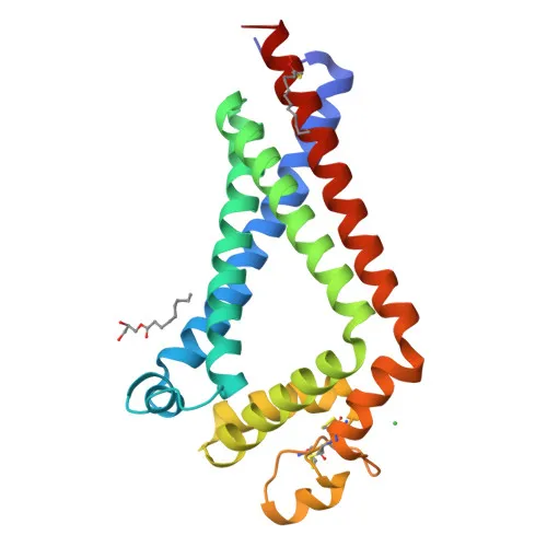
- CD9, also known as tetraspanin-29 (Tspan-29), motility-related protein (MRP-1), leukocyte antigen MIC3, and 5H9 antigen, is a member of the tetraspanin family. It is a 25 kDa integral membrane glycoprotein with four transmembrane domains and two extracellular loops.
- CD9 associates with integrins and regulates diverse processes including sperm-egg fusion, platelet activation and aggregation, and cell adhesion (PubMed:14575715, PubMed:18541721, PubMed:8478605). Related references: PMID:14575715 PMID:18541721 PMID:8478605
- In macrophages, CD9 associates with CD81 and beta-1 and beta-2 integrins, preventing macrophage fusion into multinucleated giant cells, and it similarly inhibits osteoclast formation (PubMed:12796480). Related references: PMID:12796480
- CD9 localizes to the cell membrane, is secreted in extracellular exosomes, and is expressed in platelets and a variety of hematopoietic and epithelial cells.
- CD9 is involved in cell adhesion, cell motility, and tumor metastasis (PubMed:7511626, PubMed:8478605). Related references: PMID:7511626 PMID:8478605
- The protein undergoes palmitoylation and contains disulfide bonds, which are essential for its structure and function within the tetraspanin web.
- Additional structural features include the availability of a 3D structure, and the protein is classified as a glycoprotein with direct protein sequencing evidence.
Experimental Guidance and Technical Tips
- This antibody is supplied as an conjugate, suitable for direct flow cytometry (FC) without the need for a secondary antibody. Consider titrating the antibody to determine the optimal concentration for your cell type.
- Since the immunogen is full-length recombinant human CD9 (aa 1-228), the antibody likely recognizes multiple epitopes, including extracellular domains. Cell surface staining of live cells is expected, but permeabilization may enhance staining for intracellular pools.
- For FC, use appropriate isotype controls and live/dead cell discrimination to ensure specificity. CD9 is highly expressed on platelets, so anticoagulant choice during blood collection may influence platelet activation and CD9 detection.
- CD9 is also present on exosomes; this antibody may be explored for exosome detection or immunophenotyping, but validation in the relevant sample system is required.
CamelBio: Your One-Stop Sourcing Bridge
CamelBio is your one-stop partner for IVD raw material sourcing, bridging the gap between concept and clinic. This anti-CD9 antibody supports research in immunology, oncology, and reproductive biology, and we provide a full portfolio of validated antibody pairs, bulk ancillary reagents, and customized raw material solutions to accelerate your diagnostic development.
REQUEST A QUOTE
Our professional team will reply to you within one business day. Please feel free to contact us!