Monoclonal Antibodies
Anti-Human CD2 Monoclonal Antibody () for Flow Cytometry - P06729
Item Number : CM0010788
Price varies based on specs and customizations
- Application
- FC
- Cross Reactivity
- Human
- Protein Weight
- 39kDa

Shipping:
Contact us to get shipping details Enjoy On-time Dispatch Guarantee.
Why Choose Us
Easy ordering process, quality products, and dedicated support for your business success.
Core Product Specifications and Parameters
| Parameter | Value |
|---|---|
| Product Name | Rabbit anti-Human CD2 mAb |
| Remarks/Alias | T11; SRBC; LFA-2 |
| Species | Human |
| Gene ID | 914 |
| Immunogen | Recombinant fusion protein containing a sequence corresponding to amino acids 25-209 of human CD2 (NP_001758.2). |
| Source | Rabbit |
| Category | Monoclonal Antibodies |
| Application | FC |
| Cross Reactivity | Human |
| SWISS | P06729 |
| Protein Weight | 39kDa |
| Shipping | Ice bag |
Biological Background: CD2 Function and Localization
- CD2 (T-cell surface antigen CD2) is a glycoprotein of the immunoglobulin superfamily, also designated LFA-2, T11, SRBC receptor, or Leu-5.
- It mediates cell adhesion by binding to ligands CD58 (LFA-3) and CD48, facilitating T-cell interactions with antigen-presenting cells, target cells, and other lymphocytes.
- The CD2–CD58 interaction strengthens the immunological synapse, contributing to T-cell activation, costimulation, and signal transduction.
- CD2 is expressed on T lymphocytes and natural killer (NK) cells, where it functions in both adhesion and intracellular signaling.
- Its cytoplasmic domain links to intracellular kinases and adaptor proteins, playing a critical role in T-cell triggering.
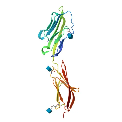
- Structurally, CD2 comprises two extracellular Ig-like domains, a single-pass transmembrane helix, and a short cytoplasmic tail containing signaling motifs.
- Subcellularly, CD2 localizes to the plasma membrane and contains disulfide bonds and N-linked glycosylation sites.
- Key post-translational modifications include glycosylation; the protein forms rosettes with sheep erythrocytes via its SRBC receptor activity.
Experimental Guidance and Technical Tips
- The immunogen used spans the extracellular domain (amino acids 25–209), targeting the ligand-binding region; thus the antibody is expected to recognize surface CD2.
- As an direct conjugate, this antibody is optimized for flow cytometry on live or fixed cells without the need for secondary reagents.
- Titrate the antibody (e.g., 0.1–5 µg per test) to determine the optimal staining concentration for your experimental system.
- Validate staining in human PBMCs or isolated T/NK cell populations; use appropriate isotype controls to set gates.
- For multicolor panels, check spectral compatibility of (excitation 491 nm, emission 518 nm) with other fluorophores.
CamelBio: Your One-Stop Sourcing Bridge
CamelBio offers comprehensive procurement solutions for IVD raw materials, supporting diagnostic developers and research laboratories with validated antibodies, bulk reagents, and rare target sourcing. For T-cell immunophenotyping and CD2-related studies, we provide directly conjugated monoclonal antibodies, such as this anti-human CD2 clone, along with ancillary reagents and custom supply options. Let us streamline your raw-material supply chain from discovery to clinical application.
REQUEST A QUOTE
Our professional team will reply to you within one business day. Please feel free to contact us!