Monoclonal Antibodies
Anti-Human CD2 Monoclonal Antibody () for Flow Cytometry - P06729
Item Number : CM0010716
Price varies based on specs and customizations
- Application
- FC
- Cross Reactivity
- Human
- Protein Weight
- 39kDa

Shipping:
Contact us to get shipping details Enjoy On-time Dispatch Guarantee.
Why Choose Us
Easy ordering process, quality products, and dedicated support for your business success.
Core Product Specifications and Parameters
| Parameter | Value |
|---|---|
| Product Name | Rabbit anti-Human CD2 mAb |
| Alias | T11, SRBC, LFA-2 |
| Species | Human |
| Gene ID | 914 |
| Immunogen | Recombinant fusion protein containing a sequence corresponding to amino acids 25-209 of human CD2 (NP_001758.2) |
| Source | Rabbit |
| Category | Monoclonal Antibodies |
| Application | FC |
| Cross Reactivity | Human |
| SWISS | P06729 |
| Protein Weight | 39kDa |
| Shipping | Ice bag |
Biological Background: CD2 Function and Localization
- CD2 (T11, SRBC, LFA-2) is a cell adhesion molecule found on the surface of T lymphocytes and natural killer cells, where it functions as a coreceptor in T-cell activation.
- It mediates intercellular adhesion by binding to its ligands CD58 (LFA-3) and CD48, facilitating the formation of immunological synapses.
- The cytoplasmic tail of CD2 is critical for signal transduction, linking extracellular binding events to intracellular signaling cascades upon T-cell stimulation.
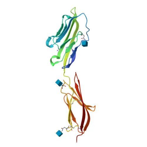
- CD2 is a transmembrane glycoprotein containing two extracellular immunoglobulin-like domains (V-type and C2-type) and a single transmembrane helix.
- While primarily expressed on T cells and NK cells, CD2 is also detected on some other leukocyte subsets, and its expression can be modulated during immune responses.
- Post-translational modifications include N-linked glycosylation, and its molecular weight is approximately 39-40 kDa.
- CD2 is the receptor for the sheep erythrocyte rosette formation, a classic assay for human T cells (hence the alias SRBC receptor).
Experimental Guidance and Technical Tips
- Titrate the antibody to determine the optimal concentration for your specific cell type and experimental setup.
- Include appropriate isotype controls and Fc receptor blocking reagents to minimize nonspecific binding.
- CD2 is expressed on T cells and NK cells; verify expression in your cellular model by co-staining with other lineage markers.
- The immunogen corresponds to the extracellular domain (aa 25-209), so the antibody likely recognizes an extracellular epitope; cell surface staining does not require permeabilization.
- Store at 2-8°C protected from light; avoid freezing, as repeated freeze-thaw cycles may reduce fluorophore stability.
- For multi-color flow cytometry panels, check spectral overlap of with other fluorophores and apply proper compensation.
CamelBio: Your One-Stop Sourcing Bridge
CamelBio offers a one-stop supply of IVD raw materials, including validated anti-CD2 antibodies for T-cell studies, alongside ancillary reagents and customized sourcing services. From monoclonal antibodies to bulk conjugates, we support every step of your diagnostic or research workflow with reliable and scalable solutions.
REQUEST A QUOTE
Our professional team will reply to you within one business day. Please feel free to contact us!