Monoclonal Antibodies
Anti-Human CD1c Monoclonal Antibody for Flow Cytometry - P29017
Item Number : CM0011374
Price varies based on specs and customizations
- Application
- FC
- Cross Reactivity
- Human
- Protein Weight
- 38kDa

Shipping:
Contact us to get shipping details Enjoy On-time Dispatch Guarantee.
Why Choose Us
Easy ordering process, quality products, and dedicated support for your business success.
Core Product Specifications and Parameters
| Parameter | Value |
|---|---|
| Product Name | Rabbit anti-Human CD1c mAb |
| Remarks/Alias | R7; CD1; CD1A; BDCA1 |
| Species | Human |
| GeneID (Human) | 911 |
| GeneID | 911 |
| Immunogen | Recombinant fusion protein containing a sequence corresponding to amino acids 19-298 of human CD1c (NP_001756.2) |
| Source | Rabbit |
| Category | Monoclonal Antibodies |
| Application | FC |
| Cross Reactivity | Human |
| SWISS | P29017 |
| Protein Weight | 38kDa |
| Shipping | Ice bag |
Biological Background: CD1c Function and Localization
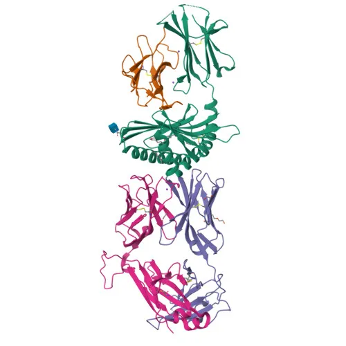
- T-cell surface glycoprotein CD1c is a member of the CD1 family of antigen-presenting molecules, encoded by the CD1C gene in humans.
- CD1c presents lipid and glycolipid antigens, both self and non-self, to T-cell receptors on natural killer T-cells, playing a central role in adaptive immunity.
- Unlike classical MHC molecules, CD1 proteins are non-polymorphic and specialize in lipid antigen presentation rather than peptide presentation.
- CD1c localizes to the cell membrane, endosome membrane, and lysosome membrane, enabling lipid antigen loading and surface display.
- Expression is detected on cortical thymocytes, certain T-cell leukemias, and various other tissues, indicating a regulated but broad tissue distribution.
- Structurally, CD1c contains an immunoglobulin domain, a transmembrane helix, and a disulfide bond; it is a glycoprotein with a mature molecular weight of approximately 38 kDa.
Experimental Guidance and Technical Tips
- The recombinant immunogen spans the extracellular region (amino acids 19–298), suggesting the antibody recognizes native CD1c. Confirm specificity using isotype controls or antigen-blocking experiments in the relevant detection system.
- For flow cytometry, titrate the antibody to determine the optimal staining concentration and achieve a suitable signal-to-noise ratio.
- has an excitation peak around 594 nm and emission around 615 nm; use appropriate laser and filter configurations (e.g., 594 nm laser, 615/20 nm filter) and protect the conjugate from prolonged light exposure.
- Consider including a viability dye and Fc receptor blocking step to minimize nonspecific binding in live-cell flow cytometry assays.
CamelBio: Your One-Stop Sourcing Bridge
CamelBio supports diagnostic manufacturers and research institutions with a comprehensive range of IVD raw materials, including validated antibody pairs, optimized monoclonal antibodies such as this anti-CD1c clone, and bulk ancillary reagents. Our sourcing network streamlines procurement for both common and rare immunological targets, covering cell-surface markers, lipid-antigen-presenting molecules, and other critical components needed for immunoassay and flow cytometry development.
REQUEST A QUOTE
Our professional team will reply to you within one business day. Please feel free to contact us!