Monoclonal Antibodies
Anti-HMOX1 (HO-1) Rabbit Monoclonal Antibody for Intracellular Flow Cytometry - P09601
Item Number : CM0008261
Price varies based on specs and customizations
- Application
- FC (intra)
- Cross Reactivity
- Human
- Protein Weight
- 33kDa

Shipping:
Contact us to get shipping details Enjoy On-time Dispatch Guarantee.
Why Choose Us
Easy ordering process, quality products, and dedicated support for your business success.
Core Product Specifications and Parameters
| Parameter | Value |
|---|---|
| Product Name | Rabbit anti-Human HMOX1 mAb |
| Remarks/Alias | HO-1; HSP32; HMOX1D; bK286B10 |
| Species | Human |
| GeneID (Human) | 3162 |
| GeneID | 3162 |
| Immunogen | Recombinant fusion protein containing a sequence corresponding to amino acids 1-265 of human Heme Oxygenase 1 (HO-1/HMOX1) (NP_002124.1) |
| Source | Rabbit |
| Category | Monoclonal Antibodies |
| Application | FC (intra) |
| Cross Reactivity | Human |
| SWISS | P09601 |
| Protein Weight | 33kDa |
| Shipping | Ice bag |
Biological Background: HMOX1 (HO-1) Function and Localization
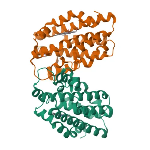
- Encodes heme oxygenase 1 (HO-1), also known as HSP32, HMOX1D, and bK286B10, a stress-inducible enzyme of ~33 kDa.
- Catalyzes the rate-limiting step in heme degradation, cleaving heme into biliverdin IXalpha, carbon monoxide (CO), and ferrous iron. Related references: PMID:11121422 PMID:19556236 PMID:7703255
- Affords cytoprotection by removing pro-apoptotic free heme, thereby inhibiting programmed cell death. Related references: PMID:20055707
- During SARS-CoV-2 infection, promotes ORF3A-mediated autophagy but is not required for ORF3A-induced reticulophagy.
- Localizes predominantly to the endoplasmic reticulum membrane.
- Expressed at higher levels in renal cancer tissue compared to normal tissue (protein level).
- Classified as an oxidoreductase with metal-binding and transmembrane domains; regulated by phosphorylation and involved in host-virus interactions.
Experimental Guidance and Technical Tips
- This antibody is recommended for intracellular flow cytometry (FC) with human samples. Consider optimizing fixation and permeabilization protocols to ensure adequate epitope accessibility.
- The immunogen spans the N-terminal 265 amino acids; thus, the antibody may recognize both full-length and N-terminal fragments of HO-1.
- HO-1 expression can be induced by oxidative stress, heme, or inflammatory stimuli—validate induction in your experimental system to confirm target upregulation.
- For multi-color flow panels, ensure the conjugate is compatible with your instrument configuration and other fluorophores.
CamelBio: Your One-Stop Sourcing Bridge
CamelBio supplies this high-quality anti-HMOX1 monoclonal antibody as part of a comprehensive IVD raw-material portfolio, supporting research in apoptosis, oxidative stress, and viral pathogenesis. We offer validated monoclonal and polyclonal antibodies, bulk ancillary reagents, and customized sourcing for rare targets—streamlining your transition from development to clinical application. Let CamelBio be your trusted partner for all IVD raw-material needs.
Product Datasheet
Anti-HMOX1 (HO-1) Rabbit Monoclonal Antibody for Intracellular Flow Cytometry - P09601
REQUEST A QUOTE
Our professional team will reply to you within one business day. Please feel free to contact us!