Polyclonal Antibodies
Anti-HLA-E Polyclonal Antibody for WB and ELISA - P13747
Item Number : CM0026397
Price varies based on specs and customizations
- Application
- WB, ELISA
- Cross Reactivity
- Human
- Protein Weight
- 40kDa

Shipping:
Contact us to get shipping details Enjoy On-time Dispatch Guarantee.
Why Choose Us
Easy ordering process, quality products, and dedicated support for your business success.
Core Product Specifications and Parameters
| Parameter | Value |
|---|---|
| Product Name | HLA-E Rabbit pAb |
| Remarks/Alias | QA1; HLA-6.2; HLA-E |
| Species | Human |
| GeneID (Human) | 3133 |
| GeneID | 3133 |
| Immunogen | Synthetic peptide corresponding to a sequence within amino acids 1-100 of human HLA-E (NP_005507.3). |
| Source | Rabbit |
| Category | Polyclonal Antibodies |
| Application | WB, ELISA |
| Cross Reactivity | Human |
| SWISS | P13747 |
| Protein Weight | 40kDa |
| Shipping | Ice bag |
Biological Background: HLA-E Function and Localization
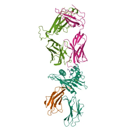
- HLA-E is a non-classical MHC class Ib molecule (HLA class I histocompatibility antigen, alpha chain E) encoded by the HLA-E gene, also known as MHC class I antigen E.
- In complex with beta-2-microglobulin, HLA-E binds nonamer self-peptides derived from signal sequences of classical MHC class I molecules (VL9 peptides) and serves as a ligand for the inhibitory NK cell receptor KLRD1-KLRC1, enabling immune monitoring of MHC class I expression and self-tolerance. Related references: PMID:17179229, PMID:18083576, PMID:18339401, PMID:37264229
- Under cellular stress, HLA-E preferentially binds stress-induced chaperone peptides, leading to loss of recognition by KLRD1-KLRC1 and impaired NK cell protection. Related references: PMID:12461076
- HLA-E presents HLA-G-derived peptides to the activating receptor KLRD1-KLRC2, contributing to adaptive NK cell generation and maternal-fetal tolerance during pregnancy. Related references: PMID:30134159, PMID:37264229, PMID:9754572
- Beyond self-peptides, HLA-E can present pathogen-derived peptides from HIV, mycobacteria, and other microbes to CD8+ cytotoxic T cells, triggering antimicrobial immune responses. Related references: PMID:16474394, PMID:20195504, PMID:30087334, PMID:34228645, PMID:35705051
- HLA-E localizes to the cell membrane and Golgi apparatus membrane; a secreted form also exists.
- It is expressed in endothelial cells of all vessel types and in lymphoid organs on B cells, T cells, monocytes, macrophages, NK cells, and megakaryocytes; also detected in secretory endometrial cells during pregnancy.
- Contains disulfide bonds, is glycosylated, phosphorylated, and has a transmembrane helix.
Experimental Guidance and Technical Tips
- The immunogen corresponds to the N-terminal region (amino acids 1-100) of human HLA-E (NP_005507.3), which includes the signal peptide. Consider that signal peptide cleavage might affect epitope availability in certain applications.
- For Western blot, the predicted band size is approximately 40 kDa. Validate detection in appropriate positive control lysates (e.g., human immune cells) and optimize exposure conditions.
- In ELISA, peptide blocking experiments can help confirm antibody specificity.
CamelBio: Your One-Stop Sourcing Bridge
CamelBio is a comprehensive IVD raw-material sourcing partner offering a wide range of validated antibodies, including the anti-HLA-E polyclonal antibody, to support immune surveillance and infectious disease research. Our one-stop platform provides optimized antibody pairs, bulk ancillary reagents, and rare target sourcing to streamline your diagnostic and assay development. Contact us to explore our full portfolio and accelerate your project from concept to clinic.
REQUEST A QUOTE
Our professional team will reply to you within one business day. Please feel free to contact us!