Polyclonal Antibodies
Anti-HLA-DRA Rabbit Polyclonal Antibody for WB, IHC-P, ELISA - P01903
Item Number : CM0016137
Price varies based on specs and customizations
- Application
- WB, IHC-P, ELISA
- Cross Reactivity
- Human, Rat
- Protein Weight
- 29kDa

Shipping:
Contact us to get shipping details Enjoy On-time Dispatch Guarantee.
Why Choose Us
Easy ordering process, quality products, and dedicated support for your business success.
Core Product Specifications and Parameters
| Parameter | Value |
|---|---|
| Product Name | HLA-DRA Rabbit pAb |
| Remarks/Alias | HLA-DRA1; HLA-DRA |
| Species | Human |
| GeneID (human) | 3122 |
| GeneID | 3122 |
| Immunogen | Recombinant fusion protein containing a sequence corresponding to amino acids 26-216 of human HLA-DRA (NP_061984.2). |
| Source | Rabbit |
| Category | Polyclonal Antibodies |
| Application | WB, IHC-P, ELISA |
| Cross Reactivity | Human, Rat |
| SWISS | P01903 |
| Protein Weight | 29kDa |
| Shipping | Ice bag |
Biological Background: HLA-DRA Function and Localization
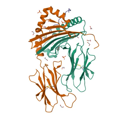
- HLA-DRA encodes the alpha chain of the HLA class II histocompatibility antigen, a heterodimeric cell surface glycoprotein that pairs with HLA-DRB to form the MHC class II complex.
- The MHCII molecule presents processed extracellular antigen peptides (10–30 amino acids) to CD4+ T helper cells, initiating adaptive immune responses such as antibody production and macrophage activation. Related references: PMID:15265931, PMID:15322540, PMID:17334368, PMID:8145819
- HLA-DRA is expressed on professional antigen-presenting cells including macrophages, dendritic cells, B cells, and thymic epithelial cells, consistent with its role in immune surveillance and T cell selection. Related references: PMID:15322540, PMID:23783831, PMID:31495665
- Subcellularly, the protein localizes to the plasma membrane and intracellular vesicular compartments—endoplasmic reticulum, endosomes, lysosomes, and autolysosomes—reflecting its trafficking for antigen loading.
- Key post-translational modifications include N-glycosylation, disulfide bond formation, and potential ubiquitination, contributing to stability and MHCII turnover.
- The protein’s structure includes a signal peptide, extracellular domains, a transmembrane helix, and a cytoplasmic tail; crystal structures of the DR heterodimer have advanced understanding of peptide binding.
- In the tumor microenvironment, HLA-DRA presents tumor-derived peptides to T cells, playing a role in anti-tumor immunity. Related references: PMID:31495665
- Autolysosomal processing of intracellular peptides by MHCII is crucial for thymic T cell selection and central immune tolerance. Related references: PMID:17182262, PMID:23783831
- The immunodominant epitope selection follows two modes: 'bind first, cut/trim later' for pathogen-derived peptides and 'cut first, bind later' for autoantigens. Related references: PMID:25413013
Experimental Guidance and Technical Tips
- The immunogen spans the extracellular domain (aa 26–216) of human HLA-DRA, enabling potential detection of both denatured and native protein conformations in applications such as WB, IHC-P, and ELISA.
- In Western blot analysis, a band around 29 kDa is expected; however, variable glycosylation may cause a shift in apparent molecular weight. Validate the band pattern in a relevant cell line or tissue lysate.
- For immunohistochemistry on paraffin-embedded sections, antigen retrieval optimization is recommended. Positive control tissues include human lymph node, spleen, or tonsil.
- Cross-reactivity with rat has been reported; for other species, check sequence homology of the immunogen region before use.
- In ELISA, the antibody may be used for capture or detection of HLA-DRA in cell culture supernatants or lysates; consider pairing with appropriate secondary antibodies and optimize coating conditions.
CamelBio: Your One-Stop Sourcing Bridge
CamelBio provides diagnostic manufacturers and researchers with a comprehensive sourcing platform for IVD raw materials, from concept to clinic. This anti-HLA-DRA antibody supports immunology research and assay development, including immune cell profiling and vaccine efficacy studies. Our portfolio also includes validated antibody pairs, bulk ancillary reagents, and rare target raw-material sourcing to streamline your diagnostic development pipeline.
Product Datasheet
Anti-HLA-DRA Rabbit Polyclonal Antibody for WB, IHC-P, ELISA - P01903
REQUEST A QUOTE
Our professional team will reply to you within one business day. Please feel free to contact us!