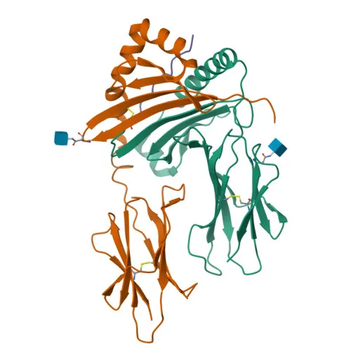
Monoclonal Antibodies
Anti-HLA-DPB1 Rabbit Monoclonal Antibody for WB, IF-P, IHC-P, ELISA - P04440
Item Number : CM0011604
Price varies based on specs and customizations
- Application
- WB, IF-P, IHC-P, ELISA
- Cross Reactivity
- Human, Mouse, Rat
- Protein Weight
- 29kDa

Shipping:
Contact us to get shipping details Enjoy On-time Dispatch Guarantee.
Why Choose Us
Easy ordering process, quality products, and dedicated support for your business success.
Core Product Specifications and Parameters
| Parameter | Value |
|---|---|
| Product Name | HLA-DPB1 Rabbit mAb |
| Remarks/Alias | DPB1; HLA-DP; HLA-DPB; HLA-DP1B |
| Species | Human |
| GeneID (Human) | 3115 |
| GeneID | 3115 |
| Immunogen | Synthetic peptide corresponding to a sequence within amino acids 1-100 of human HLA-DPB1 (NP_002112.3) |
| Source | Rabbit |
| Category | Monoclonal Antibodies |
| Application | WB, IF-P, IHC-P, ELISA |
| Cross Reactivity | Human, Mouse, Rat |
| SWISS | P04440 |
| Protein Weight | 29kDa |
| Shipping | Ice bag |
Biological Background: HLA-DPB1 Function and Localization
- HLA-DPB1 is the beta chain of the HLA class II histocompatibility antigen, a heterodimeric transmembrane glycoprotein that plays a central role in the adaptive immune response by presenting exogenous peptide antigens to CD4+ T cells. The processed peptides, typically 10–30 residues long, are loaded onto MHC class II molecules within the endosomal/lysosomal system.
- The functional MHC class II molecule is assembled as a heterononamer consisting of three alpha-beta heterodimers associated with an invariant chain (CD74) trimer. Upon entering the endosomal pathway, CD74 is progressively cleaved by proteases such as cathepsin S (CTSS) and cathepsin L (CTSL), leaving a residual CLIP peptide in the binding groove, which is then exchanged for high-affinity antigenic peptides with the assistance of the peptide editor HLA-DM.
- The antigen loading process occurs in a specialized late endosomal/lysosomal compartment, where the acidic microenvironment facilitates both proteolysis of internalized antigens and the peptide exchange reaction. In B cells, this process is further modulated by HLA-DO, which regulates the activity of HLA-DM.
- HLA-DPB1 is a member of the MHC class II beta chain gene family and maps to the HLA-DP locus on chromosome 6. Alternative names include DPB1, HLA-DP, HLA-DPB, and HLA-DP1B.
- Subcellularly, HLA-DPB1 traffics through the endoplasmic reticulum and Golgi apparatus before reaching the cell surface. It is also detected in endosomes, lysosomes, and the trans-Golgi network, reflecting its dynamic intracellular routing during peptide loading and surface expression.
- The protein is classified as a transmembrane glycoprotein with a signal peptide, a single transmembrane helix, and disulfide bonds. It undergoes N-linked glycosylation and is a member of the immunoglobulin superfamily.
- MHC class II molecules, including HLA-DPB1, are constitutively expressed on professional antigen-presenting cells such as dendritic cells, B cells, and macrophages, but can also be induced on other cell types, including intestinal epithelial cells, where they participate in mucosal immunity.
Experimental Guidance and Technical Tips
- The immunogen used to generate this antibody is a synthetic peptide spanning residues 1–100 of human HLA-DPB1, corresponding to the N‑terminal extracellular region. This region includes the signal peptide and part of the β1 domain, which is involved in peptide binding and MHC II heterodimer formation. Consider validating epitope accessibility in native versus denaturing conditions for your specific application.
- For Western blotting, a band migrating at approximately 29 kDa is expected under reducing conditions. Due to glycosylation and protein complex formation, higher molecular weight species may also be observed. Optimize blocking and wash conditions to minimize non‑specific binding in complex lysates.
- In immunofluorescence (IF-P) and immunohistochemistry (IHC-P), the antibody targets a membrane‑associated antigen. Antigen retrieval using heat‑mediated citrate buffer (pH 6.0) is often beneficial for formalin‑fixed, paraffin‑embedded sections. Include appropriate isotype controls and tissue‑specific positive controls, such as tonsil or lymph node, where HLA-DPB1 is highly expressed.
- This antibody shows cross‑reactivity with mouse and rat, indicating recognition of a conserved epitope. When using rodent samples, take into account potential differences in MHC class II cell type distribution and expression levels. Pilot experiments are recommended to confirm suitability in your model system.
- For ELISA applications, the antibody can be used as either a capture or detection reagent against recombinant or native HLA-DPB1. Pairing with an anti‑HLA‑DP alpha chain antibody may help ensure detection of the intact heterodimer. Validate the assay in the relevant sample matrix to rule out interference from soluble HLA or cross‑reactive MHC class I molecules.
CamelBio: Your One-Stop Sourcing Bridge
CamelBio serves as a comprehensive procurement partner for diagnostic manufacturers and research laboratories, providing a reliable supply of premium raw materials, including the anti-HLA-DPB1 antibody for MHC class II antigen presentation research. Through flexible sourcing models, we offer validated antibody pairs, optimized monoclonal and polyclonal antibodies, bulk ancillary reagents, and custom rare‑target raw‑material sourcing to accelerate your IVD development pipeline from concept to clinic. Contact our team to discuss how our one‑stop services can meet your specific reagent needs and project timelines.
Product Datasheet
Anti-HLA-DPB1 Rabbit Monoclonal Antibody for WB, IF-P, IHC-P, ELISA - P04440
REQUEST A QUOTE
Our professional team will reply to you within one business day. Please feel free to contact us!