Monoclonal Antibodies
Anti-Histone H2B (testis specific) Monoclonal Antibody for WB, IP, IHC - Q96A08
Item Number : CM0006770
Price varies based on specs and customizations
- Application
- WB, IP, IF-P, IHC-P, ELISA
- Cross Reactivity
- Human, Mouse, Rat, Other (Wide Range Predicted)
- Protein Weight
- 14kDa

Shipping:
Contact us to get shipping details Enjoy On-time Dispatch Guarantee.
Why Choose Us
Easy ordering process, quality products, and dedicated support for your business success.
Core Product Specifications and Parameters
| Parameter | Value |
|---|---|
| Product Name | Histone H2B (testis specific) Rabbit mAb |
| Remarks/Alias | STBP; TH2B; H2BFU; TSH2B; hTSH2B; TSH2B.1; HIST1H2BA; bA317E16.3 |
| Species | Human |
| GeneID (human) | 255626 |
| GeneID | 255626 |
| Immunogen | Synthetic peptide corresponding to amino acids 1-100 of human Histone H2B (testis specific) (NP_733759.1) |
| Source | Rabbit |
| Category | Monoclonal Antibodies |
| Application | WB, IP, IF-P, IHC-P, ELISA |
| Cross Reactivity | Human, Mouse, Rat, Other (Wide Range Predicted) |
| SWISS | Q96A08 |
| Protein Weight | 14kDa |
| Shipping | Ice bag |
Biological Background: Histone H2B type 1-A Function and Localization
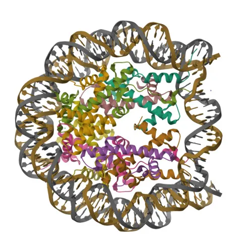
- Histone H2B type 1-A (H2BC1, also known as TSH2B.1/hTSH2B) is a testis-specific variant histone that replaces classical histone H2B during spermatogenesis.
- It specifically directs the transformation of dissociating nucleosomes to protamine in male germ cells, acting as a nucleosome dissociating factor to create dynamic chromatin facilitating large-scale histone exchange (By similarity).
- As a core component of the nucleosome, it wraps and compacts DNA into chromatin, playing a central role in transcription regulation, DNA repair, DNA replication, and chromosomal stability (By similarity).
- DNA accessibility is regulated through a complex set of post-translational modifications (histone code) and nucleosome remodeling (By similarity).
- Histone H2B type 1-A is also expressed in some fat cells; its function and specific post-translational modifications in these cells remain unclear (PubMed:21249133). Related references: PMID:21249133
- Subcellular localization is restricted to the nucleus and chromosomes.
- Known post-translational modifications include acetylation, methylation, phosphorylation, isopeptide bond formation, and ubiquitin-like conjugation.
- While predominantly expressed in testis and mature sperm (at protein level), it is also detected in certain fat cells.
Experimental Guidance and Technical Tips
- The immunogen corresponds to the N-terminal region (aa 1-100) of human Histone H2B type 1-A; therefore, the antibody likely detects an epitope in this region, which is conserved across the predicted cross-reactive species.
- For Western blot, expect a band at ~14 kDa; validate in a relevant sample system such as human testis lysate or other tissues expressing this variant.
- For immunohistochemistry (IHC-P) and immunofluorescence (IF-P), consider testis tissue sections where expression is highest; optimization of antigen retrieval may be required.
- For immunoprecipitation (IP), use a nuclear extract from testis or cells expressing the target; confirm interactions with protamine exchange factors if relevant.
- As this histone variant is also found in some fat cells, researchers may detect bands or staining in adipose tissue or adipocyte cultures; consider using appropriate controls to verify specificity.
CamelBio: Your One-Stop Sourcing Bridge
CamelBio serves as a comprehensive sourcing partner for IVD raw materials, connecting diagnostic manufacturers and researchers with high-quality antibodies, antigens, and ancillary reagents. This testis-specific Histone H2B antibody supports studies in chromatin dynamics and male fertility research. Leverage CamelBio’s supply network for validated antibody pairs, bulk reagent orders, and custom sourcing of rare-target raw materials to accelerate assay development.
REQUEST A QUOTE
Our professional team will reply to you within one business day. Please feel free to contact us!