Polyclonal Antibodies
Anti-HIST1H3D Polyclonal Antibody for WB, ELISA - Q16695 / P68431
Item Number : CM0023055
Price varies based on specs and customizations
- Application
- WB, ELISA
- Cross Reactivity
- Human, Mouse, Rat, Other (Wide Range Predicted)
- Protein Weight
- 15KDa

Shipping:
Contact us to get shipping details Enjoy On-time Dispatch Guarantee.
Why Choose Us
Easy ordering process, quality products, and dedicated support for your business success.
Core Product Specifications and Parameters
| Parameter | Value |
|---|---|
| Product Name | HIST1H3D Rabbit pAb |
| Remarks/Alias | H3.4; H3/g; H3FT; H3t; HIST3H3; Histone H3; HIST1H3A; HIST1H3D |
| Species | Human |
| Gene ID (Human) | 8290/8350 |
| Gene ID | 8290/8350 |
| Immunogen | Recombinant fusion protein containing a sequence corresponding to amino acids 1-136 of human HIST1H3D (NP_003520.1). |
| Source | Rabbit |
| Category | Polyclonal Antibodies |
| Application | WB, ELISA |
| Cross Reactivity | Human, Mouse, Rat, Other (Wide Range Predicted) |
| SWISS | Q16695/P68431 |
| Protein Weight | 15KDa |
| Shipping | Ice bag |
Biological Background: Histone H3.1t Function and Localization
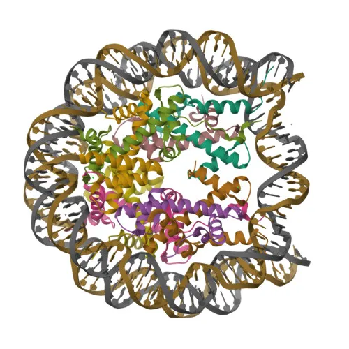
- Histone H3.1t (also known as H3/g or H3.4) is a core component of the nucleosome, wrapping and compacting DNA into chromatin to regulate accessibility for transcription, replication, and repair.
- It plays a central role in maintaining chromosomal stability and is subject to a complex set of post-translational modifications, collectively referred to as the histone code, which includes acetylation, methylation, phosphorylation, and ubiquitination.
- Encoded by the H3-4 gene (also designated H3FT or HIST3H3), this replication-independent histone variant is primarily expressed in testicular cells, suggesting a specialized function in spermatogenesis.
- Subcellularly, Histone H3.1t localizes to the nucleus and is specifically associated with chromosomes, where it forms nucleosome core particles.
- Key post-translational modifications include acetylation, methylation, phosphorylation, citrullination, ADP-ribosylation, hydroxylation, and ubiquitination, which regulate DNA binding, chromatin structure, and gene expression.
Experimental Guidance and Technical Tips
- For Western blot (WB), the predicted molecular weight of Histone H3.1t is approximately 15 kDa. Optimize membrane pore size and transfer conditions for low-molecular-weight proteins.
- For ELISA, use the recombinant immunogen as a positive control to validate antibody binding specificity.
- Because histone proteins are highly conserved across species, the predicted wide cross-reactivity (human, mouse, rat, and others) should be verified in the target species lysates or cell lines.
- Consider using acid extraction or histone-enriched nuclear fractions to improve detection sensitivity in WB, as histones are highly abundant but may require specific extraction methods.
CamelBio: Your One-Stop Sourcing Bridge
CamelBio provides diagnostic manufacturers, labs, and research institutes with a reliable one-stop sourcing platform for IVD raw materials. For chromatin biology and histone modification research, we offer validated antibody pairs, optimized polyclonal antibodies, and bulk ancillary reagents to support your assay development. Whether you need rare-target antibodies or large-scale production, CamelBio bridges your project from concept to clinical application.
REQUEST A QUOTE
Our professional team will reply to you within one business day. Please feel free to contact us!