Monoclonal Antibodies
Anti-HIBADH Monoclonal Antibody for WB, IHC-P, IF/ICC, ELISA - P31937
Item Number : CM0007630
Price varies based on specs and customizations
- Application
- WB, IHC-P, IF/ICC, ELISA
- Cross Reactivity
- Human, Mouse, Rat
- Protein Weight
- 35kDa

Shipping:
Contact us to get shipping details Enjoy On-time Dispatch Guarantee.
Why Choose Us
Easy ordering process, quality products, and dedicated support for your business success.
Core Product Specifications and Parameters
| Parameter | Specification |
|---|---|
| Product Name | HIBADH Rabbit mAb |
| Remarks/Alias | NS5ATP1; HIBADH |
| Species | Human |
| GeneID (Human) | 11112 |
| GeneID | 11112 |
| Immunogen | Recombinant fusion protein containing a sequence corresponding to amino acids 37-336 of human HIBADH (NP_689953.1) |
| Source | Rabbit |
| Category | Monoclonal Antibodies |
| Application | WB, IHC-P, IF/ICC, ELISA |
| Cross Reactivity | Human, Mouse, Rat |
| SWISS | P31937 |
| Protein Weight | 35kDa |
| Shipping | Ice bag |
Biological Background: HIBADH Function and Localization
- HIBADH (3-hydroxyisobutyrate dehydrogenase) is a mitochondrial enzyme belonging to the oxidoreductase family, utilizing NAD as a cofactor.
- It plays a key role in the catabolism of branched-chain amino acids (valine, leucine, isoleucine), specifically in the conversion of 3-hydroxyisobutyrate to methylmalonate semialdehyde.
- The protein is localized to the mitochondrion, as indicated by its mitochondrial transit peptide, which is cleaved to yield the mature enzyme.
- Post-translational modifications include acetylation, which may regulate its enzymatic activity or stability.
- HIBADH expression has been detected in skin fibroblasts; its expression in other tissues may be inferred from its metabolic role.
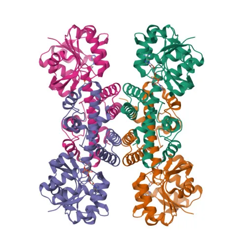
- Structural studies have provided a 3D-structure for HIBADH, aiding in understanding its catalytic mechanism and substrate recognition.
- The protein has been identified through direct protein sequencing and proteomics approaches, confirming its presence in human mitochondria.
Experimental Guidance and Technical Tips
- The immunogen corresponds to amino acids 37-336 of human HIBADH (NP_689953.1), which excludes the predicted mitochondrial transit peptide (approximately the first 36 amino acids); the antibody is therefore likely to recognize the mature mitochondrial form.
- For Western blot (WB), a band at approximately 35 kDa is expected (consistent with UniProt molecular weight of 35.3 kDa). Confirm specificity by using appropriate positive and negative controls (e.g., HIBADH-overexpressing cells or tissues with known expression).
- In immunohistochemistry (IHC-P) and immunofluorescence (IF/ICC), mitochondrial staining patterns should be observed. Co-localization with mitochondrial markers can help validate specificity.
- The antibody cross-reacts with human, mouse, and rat HIBADH; when using rodent samples, verify sequence homology in the immunogen region to ensure comparable reactivity.
- For ELISA applications, consider using the recombinant immunogen as a positive control and titrate the antibody concentration for optimal signal-to-noise ratio.
- To minimize non-specific binding, suitable blocking buffers (e.g., 5% non-fat milk or BSA) and appropriate dilution optimization are recommended.
CamelBio: Your One-Stop Sourcing Bridge
CamelBio supports diagnostic manufacturers and researchers by providing validated antibody pairs, optimized monoclonal/polyclonal antibodies, bulk ancillary reagents, and rare target raw-material sourcing. For metabolic and mitochondrial disease research, CamelBio offers HIBADH-specific reagents and customizable raw-material solutions to accelerate assay development. Our one-stop supply chain ensures seamless transition from concept to clinical application.
Product Datasheet
Anti-HIBADH Monoclonal Antibody for WB, IHC-P, IF/ICC, ELISA - P31937
REQUEST A QUOTE
Our professional team will reply to you within one business day. Please feel free to contact us!