Monoclonal Antibodies
Anti-HFE Monoclonal Antibody for WB, ELISA - Q30201
Item Number : CM0002416
Price varies based on specs and customizations
- Application
- WB, ELISA
- Cross Reactivity
- Human
- Protein Weight
- 40kDa

Shipping:
Contact us to get shipping details Enjoy On-time Dispatch Guarantee.
Why Choose Us
Easy ordering process, quality products, and dedicated support for your business success.
Core Product Specifications and Parameters
| Parameter | Value |
|---|---|
| Product Name | HFE Rabbit mAb |
| Remarks/Alias | HH; HFE1; HLA-H; MVCD7; TFQTL2; HFE |
| Species | Human |
| GeneID (human) | 3077 |
| GeneID | 3077 |
| Immunogen | Synthetic Peptide|A synthetic peptide corresponding to a sequence within amino acids 249-348 of human HFE (Q30201). |
| Source | Rabbit |
| Category | Monoclonal Antibodies |
| Application | WB, ELISA |
| Cross Reactivity | Human |
| SWISS | Q30201 |
| Protein Weight | 40kDa |
| Shipping | Ice bag |
Biological Background: HFE Function and Localization
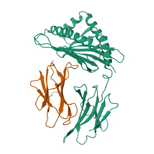
- HFE (Hereditary Hemochromatosis Protein), also referred to as HLA-H, is a membrane protein that binds to the transferrin receptor (TFR) and reduces its affinity for iron-loaded transferrin, thereby regulating iron uptake.
- The gene encoding HFE belongs to the MHC class I family; mutations in HFE are associated with hereditary hemochromatosis, a disorder of iron overload, consistent with its classification as a disease variant protein.
- According to UniProt, HFE is expressed in all tissues tested except the brain, indicating a broad but not ubiquitous distribution.
- Subcellularly, HFE localizes to the cell membrane, where it interacts with TFR and other iron-related proteins to control systemic iron homeostasis.
- Alternative splicing of HFE produces multiple isoforms, which may have distinct tissue distributions and functional roles; the full-length membrane-bound form is the primary species.
- Post-translational modifications include glycosylation and disulfide bond formation, which are critical for proper folding and cell surface expression.
Experimental Guidance and Technical Tips
- The immunogen corresponds to a synthetic peptide spanning amino acids 249–348 of human HFE, a region that includes part of the extracellular domain, the transmembrane helix, and the cytoplasmic tail. This design may enable detection of both full-length and certain truncated forms in denaturing Western blot.
- For Western blotting, consider using total protein lysates from HFE-expressing tissues such as liver or placenta (excluding brain, where HFE is absent). Optimize blocking and antibody dilution conditions for your specific sample type.
- For ELISA applications, the antibody’s performance may depend on the assay format (direct, indirect, or sandwich). Validate the clone in your target sample matrix and consider pairing with a capture antibody that recognizes a distinct epitope.
- As HFE is a membrane protein, ensure proper solubilization and denaturation during sample preparation to expose the epitope, particularly if the immunogen is partially intracellular.
- Cross-reactivity is reported for human HFE; for use with other species, sequence homology should be verified.
CamelBio: Your One-Stop Sourcing Bridge
CamelBio bridges the gap between diagnostic developers and critical raw materials, offering this validated anti-HFE monoclonal antibody as part of a comprehensive portfolio for iron metabolism and hemochromatosis research. In addition to ready-to-use antibodies, we provide bulk sourcing of ancillary reagents and custom procurement for rare targets, supporting your workflow from early concept to clinical application. Contact us to discuss your specific antibody pair and reagent needs.
REQUEST A QUOTE
Our professional team will reply to you within one business day. Please feel free to contact us!