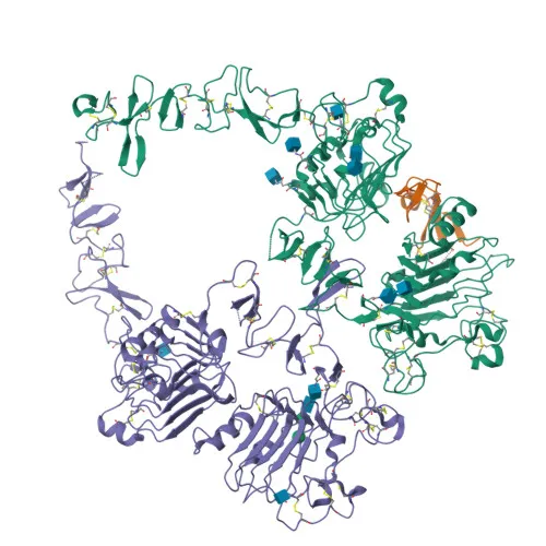
Monoclonal Antibodies
Anti-HER2/ErbB2 Monoclonal Antibody for Flow Cytometry - P04626
Item Number : CM0008796
Price varies based on specs and customizations
- Application
- FC
- Cross Reactivity
- Human
- Protein Weight
- 138kDa

Shipping:
Contact us to get shipping details Enjoy On-time Dispatch Guarantee.
Why Choose Us
Easy ordering process, quality products, and dedicated support for your business success.
Core Product Specifications and Parameters
| Parameter | Value |
|---|---|
| Product Name | Rabbit anti-Human HER2/ErbB2 mAb |
| Remarks/Alias | NEU; NGL; HER2; TKR1; CD340; HER-2; VSCN2; MLN 19; MLN-19; c-ERB2; c-ERB-2; HER-2/neu; p185(erbB2) |
| Species | Human |
| GeneID (Human) | 2064 |
| GeneID | 2064 |
| Immunogen | Recombinant protein: Recombinant fusion protein containing a sequence corresponding to amino acids 23-652 of human HER2/ErbB2 (NP_004439.2) |
| Source | Rabbit |
| Category | Monoclonal Antibodies |
| Application | FC |
| Cross Reactivity | Human |
| SWISS | P04626 |
| Protein Weight | 138kDa |
| Shipping | Ice bag |
Biological Background: HER2/ErbB2 Function and Localization
- Protein Identity and Isoforms: Receptor tyrosine-protein kinase erbB-2, also known as HER2, Neu, CD340, or p185erbB2. Encoded by the ERBB2 gene, it functions as a critical cell surface receptor in signal transduction pathways controlling cell growth and differentiation.
- Core Nucleosome/Chromatin Function: Not directly a chromatin protein, but HER2 signaling influences gene expression programs. It is an essential component of neuregulin-receptor complexes, though neuregulins do not bind it directly; instead, it requires a coreceptor for ligand interaction. GP30 is a potential ligand.
- Microtubule Regulation: Upon activation, HER2 triggers the MEMO1-RHOA-DIAPH1 pathway, phosphorylating and inhibiting GSK3B. This prevents phosphorylation of APC and CLASP2, enabling their membrane association and subsequent MACF1 localization, which captures and stabilizes peripheral microtubules.
- Nuclear Functions: Translocates to the nucleus where it acts as a transcriptional regulator. Binds to the 5'-TCAAATTC-3' sequence in the PTGS2/COX-2 promoter to activate transcription. Also participates in STAT3- and SRC-dependent CDKN1A transcription and enhances rRNA synthesis via RNA Pol I, promoting protein translation and cell growth.
- Subcellular Localization: Found at the cell membrane, ruffles, early endosomes, cytoplasm (perinuclear region), and nucleus. This dynamic distribution reflects its diverse roles in membrane-proximal signaling and nuclear gene regulation.
- Tissue Specificity and Disease Association: Expressed in various tumor tissues, including primary breast tumors, and cancers of the small bowel, esophagus, kidney, and oral cavity. Amplification/overexpression is a hallmark of aggressive cancers, making HER2 a key diagnostic and therapeutic target.
- Key Post-translational Modifications and Keywords: Contains a signal peptide, a large extracellular domain, a single-pass transmembrane helix, and an intracellular tyrosine kinase domain. Heavily glycosylated with disulfide bonds. Autophosphorylation on tyrosine residues is critical for kinase activity. Keywords include ATP-binding, kinase, transferase, glycoprotein, phosphoprotein, disease variant, proto-oncogene.
Experimental Guidance and Technical Tips
- This antibody was raised against a recombinant protein corresponding to the extracellular domain (amino acids 23‑652) of human HER2, making it suitable for detecting cell surface HER2 by flow cytometry (FC) without permeabilization. Consider titrating the antibody to optimize signal-to-noise ratios for your specific cell model.
- For positive controls, use HER2‑overexpressing cell lines such as SK‑BR‑3 or BT‑474. Validate staining with an appropriate isotype control to confirm specificity.
- If intracellular or total HER2 detection is required, note that fixation and permeabilization may alter epitope accessibility. Validate the antibody in your assay conditions and consider live‑cell staining for surface‑only targets.
- Cross‑reactivity with species other than human has not been tested. If working with non‑human samples, confirm reactivity experimentally.
- Always include a viability dye to exclude dead cells, which can increase non‑specific binding.
CamelBio: Your One-Stop Sourcing Bridge
CamelBio empowers diagnostic manufacturers, research laboratories, and institutes with a comprehensive, one-stop portfolio of IVD raw materials, technical services, and consulting—supporting every stage from concept to clinic. For HER2/ErbB2‑focused projects, we offer validated antibody pairs, optimized monoclonal and polyclonal antibodies, bulk ancillary reagents, and rare‑target raw material sourcing. Whether you are developing cancer biomarker assays or seeking reliable HER2 detection reagents, our integrated supply chain and expertise help accelerate your diagnostic development pipeline.
REQUEST A QUOTE
Our professional team will reply to you within one business day. Please feel free to contact us!