Polyclonal Antibodies
Anti-HCoV-HKU1 Spike S1 Polyclonal Antibody for WB, ELISA - Q5MQD0
Item Number : CM0020423
Price varies based on specs and customizations
- Application
- WB, ELISA
- Cross Reactivity
- HCoV-HKU1
- Protein Weight
- 152 kDa

Shipping:
Contact us to get shipping details Enjoy On-time Dispatch Guarantee.
Why Choose Us
Easy ordering process, quality products, and dedicated support for your business success.
Core Product Specifications and Parameters
| Parameter | Value |
|---|---|
| Product Name | HCoV-HKU1 Spike S1 Rabbit pAb |
| Species | Human coronavirus HKU1 |
| Gene ID | 3200426 |
| Immunogen | A synthetic peptide corresponding to a sequence within amino acids 100-200 of coronavirus Spike S1 (YP_173238.1) |
| Source | Rabbit |
| Category | Polyclonal Antibodies |
| Application | WB, ELISA |
| Cross Reactivity | HCoV-HKU1 |
| SWISS | Q5MQD0 |
| Protein Weight | 152 kDa |
| Shipping | Ice bag |
Biological Background: HCoV-HKU1 Spike S1 Function and Localization
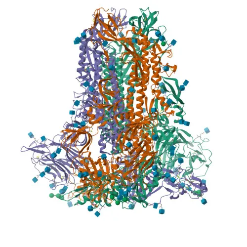
- Spike glycoprotein (S), also known as E2 or peplomer protein, is the major envelope protein of HCoV-HKU1 responsible for host cell attachment and entry.
- The S protein is cleaved into S1 and S2 subunits; the S1 subunit contains the receptor-binding domain (RBD) that interacts with host receptors to initiate infection.
- Following receptor binding, the S2 subunit undergoes conformational changes, exposing the fusion peptide and mediating fusion of the viral and cellular membranes as a class I fusion protein.
- Heptad repeat regions in S2 assemble into a trimer-of-hairpins structure, bringing the fusion peptide close to the target membrane, which drives membrane apposition and fusion.
- The fusion peptide is unmasked after S2 cleavage, which occurs upon virus endocytosis, facilitating viral entry into the host cytoplasm.
- Subcellular localization includes the virion membrane, host cell membrane, and host endoplasmic reticulum-Golgi intermediate compartment membrane.
- The protein undergoes key post-translational modifications, including disulfide bond formation, glycosylation, and palmitoylation (lipoprotein), which are critical for its structural integrity and function.
Experimental Guidance and Technical Tips
- In western blot, a band of approximately 152 kDa corresponding to the full-length Spike protein is expected; reducing conditions may be required for optimal detection.
- To confirm specificity, consider using lysates from HCoV-HKU1-infected cells or recombinant S1 protein as positive controls, and include uninfected or heterologous coronavirus lysates as negative controls.
- For ELISA, this antibody can be used for direct detection or as a capture antibody; pairing with a different clone or detection method may improve assay sensitivity.
- Note that cross-reactivity has been validated only for HCoV-HKU1; performance with other coronaviruses should be independently verified.
CamelBio: Your One-Stop Sourcing Bridge
CamelBio provides diagnostic manufacturers and research institutions with streamlined access to essential IVD raw materials. For coronavirus research programs, we offer validated antibody pairs, optimized monoclonal and polyclonal antibodies, bulk ancillary reagents, and customized sourcing of rare-target raw materials—all from a single point of contact. Our comprehensive supply chain supports your workflow from early-stage concept through clinical application.
REQUEST A QUOTE
Our professional team will reply to you within one business day. Please feel free to contact us!