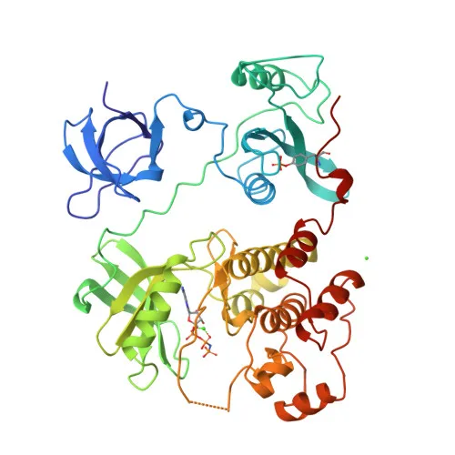
Polyclonal Antibodies
Anti-HCK Rabbit Polyclonal Antibody for WB, IF/ICC, ELISA - P08631
Item Number : CM0022186
Price varies based on specs and customizations
- Application
- WB, IF/ICC, ELISA
- Cross Reactivity
- Human
- Protein Weight
- 60kDa

Shipping:
Contact us to get shipping details Enjoy On-time Dispatch Guarantee.
Why Choose Us
Easy ordering process, quality products, and dedicated support for your business success.
Core Product Specifications and Parameters
| Parameter | Specification |
|---|---|
| Product Name | HCK Rabbit pAb |
| Remarks/Alias | JTK9, AIPCV, p59Hck, p61Hck, HCK |
| Species | Human |
| GeneID (human) | 3055 |
| GeneID | 3055 |
| Immunogen | Recombinant fusion protein containing a sequence corresponding to amino acids 1-220 of human HCK (NP_001165600.1) |
| Source | Rabbit |
| Category | Polyclonal Antibodies |
| Application | WB, IF/ICC, ELISA |
| Cross Reactivity | Human |
| SWISS | P08631 |
| Protein Weight | 60kDa |
| Shipping | Ice bag |
Biological Background: HCK Function and Localization
- Tyrosine-protein kinase HCK is a non-receptor kinase predominantly expressed in hematopoietic cells, particularly myeloid and B-lymphoid lineages; alternative splicing and initiation produce isoforms p59Hck and p61Hck.
- Acts as a key signal transducer downstream of various receptors (Fc receptors, cytokine receptors, integrins) to regulate innate immune responses, including phagocytosis, degranulation, respiratory burst, cell adhesion, migration, and survival.
- Mediates phosphorylation of multiple substrates such as CBL, VAV1, STAT5B, and WAS, thereby controlling actin cytoskeleton reorganization, podosome formation, and cell protrusion.
- Localizes to multiple subcellular compartments including plasma membrane, caveolae, focal adhesions, podosomes, cytoplasm, lysosomes, secretory vesicles, Golgi, and nucleus, reflecting its dynamic role in signal transduction.
- Highly expressed in granulocytes, and detected in monocytes and neutrophils; also found in tonsil tissue.
- Contains SH2 and SH3 domains for protein-protein interactions and an ATP-binding kinase domain; subject to myristoylation and palmitoylation for membrane targeting.
- Identified as a proto-oncogene and reference proteome member; also undergoes ubiquitin-like conjugation.
Experimental Guidance and Technical Tips
- This polyclonal antibody was raised against the N-terminal region (aa 1-220) of human HCK, which may detect both p59 and p61 isoforms; consider isoform-specific validation if needed.
- For WB, a band around 60 kDa is expected; post-translational modifications may cause minor electrophoretic shifts.
- Recommended applications include WB, IF/ICC, and ELISA; optimize dilutions and appropriate controls for each assay system.
- Reported cross-reactivity is for human; for other species, check sequence homology in the immunogen region before use.
CamelBio: Your One-Stop Sourcing Bridge
CamelBio offers diagnostic manufacturers and researchers a single gateway to high-quality IVD raw materials, including validated polyclonal antibodies like HCK Rabbit pAb for studies in innate immunity and hematopoietic cell signaling. Our portfolio spans optimized antibody pairs, bulk ancillary reagents, and customized sourcing for rare targets, streamlining your development from concept to clinic.
REQUEST A QUOTE
Our professional team will reply to you within one business day. Please feel free to contact us!