Polyclonal Antibodies
Anti-HADH Rabbit Polyclonal Antibody for WB, IHC-P, ELISA - Q16836
Item Number : CM0013296
Price varies based on specs and customizations
- Application
- WB, IHC-P, ELISA
- Cross Reactivity
- Human, Mouse, Rat
- Protein Weight
- 34kDa

Shipping:
Contact us to get shipping details Enjoy On-time Dispatch Guarantee.
Why Choose Us
Easy ordering process, quality products, and dedicated support for your business success.
Core Product Specifications and Parameters
| Parameter | Value |
|---|---|
| Product Name | HADH Rabbit pAb |
| Remarks/Alias | HAD; HCDH; HHF4; HADH1; HADHSC; MSCHAD; HADH |
| Species | Human |
| GeneID (Human) | 3033 |
| GeneID | 3033 |
| Immunogen | Recombinant fusion protein containing a sequence corresponding to amino acids 1-314 of human HADH (NP_005318.3). |
| Source | Rabbit |
| Category | Polyclonal Antibodies |
| Application | WB, IHC-P, ELISA |
| Cross Reactivity | Human, Mouse, Rat |
| SWISS | Q16836 |
| Protein Weight | 34kDa |
| Shipping | Ice bag |
Biological Background: HADH Function and Localization
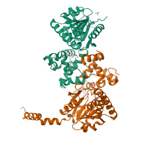
- HADH (Hydroxyacyl-coenzyme A dehydrogenase, mitochondrial) catalyzes the third step of the mitochondrial beta-oxidation cycle for medium and short-chain 3-hydroxy fatty acyl-CoAs (C4 to C10), essential for energy production from fatty acids (PubMed:10231530, PubMed:11489939, PubMed:16725361). Related references: PMID:10231530 PMID:11489939 PMID:16725361
- Also designated as medium and short-chain L-3-hydroxyacyl-coenzyme A dehydrogenase or short-chain 3-hydroxyacyl-CoA dehydrogenase (SCHAD), the protein plays a central role in lipid metabolism.
- Beyond beta-oxidation, HADH negatively regulates insulin secretion by inhibiting glutamate dehydrogenase 1 (GLUD1) activation, linking fatty acid metabolism to glucose homeostasis (By similarity).
- The enzyme contributes to the maintenance of normal spermatogenesis by reducing fatty acid accumulation in the testes, highlighting a specialized role in reproductive tissues (By similarity).
- HADH localizes to the mitochondrial matrix, where it functions as an NAD-dependent oxidoreductase.
- Post-translational modifications include acetylation and hydroxylation, which may modulate enzymatic activity or stability.
- Tissue expression is highest in liver, kidney, pancreas, heart, and skeletal muscle, consistent with tissues having high energy demands or specialized metabolic functions.
- Mutations in HADH are associated with disease variants such as hyperinsulinemic hypoglycemia, emphasizing its clinical relevance in metabolic disorders.
Experimental Guidance and Technical Tips
- The immunogen encompasses full-length human HADH (aa 1–314); while it may cross-react with mouse and rat orthologs, species-specific validation is recommended.
- For western blotting, anticipate a specific band near 34 kDa; optimize gel percentage and transfer conditions for low-molecular-weight proteins.
- In IHC-paraffin applications, consider testing different antigen retrieval methods and antibody dilutions on positive control tissues such as human liver or kidney.
- For ELISA, titrate the antibody against recombinant HADH or native protein in the relevant sample matrix to establish optimal working concentrations.
CamelBio: Your One-Stop Sourcing Bridge
CamelBio serves as a strategic one-stop partner for diagnostic manufacturers and research institutes, providing validated IVD raw materials like this anti-HADH polyclonal antibody for metabolic disease and mitochondrial research. Our comprehensive catalogue includes validated antibody pairs, optimized monoclonal and polyclonal antibodies, bulk ancillary reagents, and rare-target sourcing support to accelerate your assay development pipeline.
REQUEST A QUOTE
Our professional team will reply to you within one business day. Please feel free to contact us!