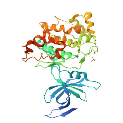
Monoclonal Antibodies
Anti-GSK3β Monoclonal Antibody for Flow Cytometry (Intracellular) - P49841
Item Number : CM0005589
Price varies based on specs and customizations
- Application
- FC (intra)
- Cross Reactivity
- Human
- Protein Weight
- 47kDa

Shipping:
Contact us to get shipping details Enjoy On-time Dispatch Guarantee.
Why Choose Us
Easy ordering process, quality products, and dedicated support for your business success.
Core Product Specifications and Parameters
| Parameter | Value |
|---|---|
| Product Name | Rabbit anti-Human GSK3β mAb |
| Remarks/Alias | GSK3B; glycogen synthase kinase-3 beta; GSK3β |
| Species | Human |
| GeneID (human) | 2932 |
| GeneID | 2932 |
| Immunogen | Synthetic peptide corresponding to a sequence within amino acids 321-420 of human GSK3β (NP_001139628.1). |
| Source | Rabbit |
| Category | Monoclonal Antibodies |
| Application | FC (intra) |
| Cross Reactivity | Human |
| SWISS | P49841 |
| Protein Weight | 47kDa |
| Shipping | Ice bag |
Biological Background: GSK3β Function and Localization
- GSK3β is a constitutively active serine/threonine kinase that acts as a negative regulator in hormonal control of glucose homeostasis, Wnt signaling, and regulation of transcription factors and microtubules, by phosphorylating substrates such as glycogen synthase, β-catenin, and Tau. Related references: PMID:11430833 PMID:12554650 PMID:14690523
- It requires primed phosphorylation of most substrates; in skeletal muscle, it phosphorylates and inhibits glycogen synthase (GYS1), regulating glycogen synthesis and potentially contributing to insulin resistance. Related references: PMID:8397507 PMID:11430833 PMID:16484495
- In Wnt signaling, GSK3β forms a complex with APC, AXIN1, and β-catenin, phosphorylating β-catenin to promote its ubiquitin-mediated degradation. Related references: PMID:12554650
- GSK3β phosphorylates Tau (MAPT) at Thr-548, reducing microtubule binding; Tau is a major component of neurofibrillary tangles in Alzheimer disease, and GSK3β colocalizes with Tau and PKR in Alzheimer brains. Related references: PMID:14690523
- It regulates the circadian clock by phosphorylating core clock proteins BMAL1, CLOCK, and PER2, targeting them for proteasomal degradation. Related references: PMID:19946213 PMID:28903391
- GSK3β localizes to cytoplasm, nucleus, and cell membrane; expressed highly in testis, thymus, prostate, ovary, and weakly in lung, brain, kidney. The gene undergoes alternative splicing, and the protein is modified by phosphorylation, ADP-ribosylation, and palmitoylation.
Experimental Guidance and Technical Tips
- The antibody targets an intracellular epitope; fixation and permeabilization are required for intracellular flow cytometry. Optimize permeabilization conditions for your cell type.
- Validate GSK3β expression in relevant cell lines (e.g., HEK293, HeLa) using positive and negative controls to confirm staining specificity.
- Use appropriate isotype controls and titrate the conjugated antibody to minimize background and optimize signal-to-noise ratio.
- Since the immunogen spans amino acids 321-420 near the C-terminus, ensure epitope accessibility is not masked by protein interactions or modifications.
- Check compensation settings if using in multi-color panels; reference dye spectral overlap guides.
- No cross-reactivity data is provided; verify compatibility with your target species or cell line of interest.
CamelBio: Your One-Stop Sourcing Bridge
CamelBio is a dedicated IVD raw-material partner, providing streamlined sourcing of optimized antibodies, validated antibody pairs, and bulk ancillary reagents to support diagnostic development. Our technical team facilitates access to rare targets and offers consultation to help you move from concept to clinical application smoothly. Explore this GSK3β antibody for intracellular flow cytometry in metabolic, neurodegenerative, and oncology research areas.
Product Datasheet
Anti-GSK3β Monoclonal Antibody for Flow Cytometry (Intracellular) - P49841
REQUEST A QUOTE
Our professional team will reply to you within one business day. Please feel free to contact us!