Polyclonal Antibodies
Anti-GRK5 Polyclonal Antibody for WB, IHC-P, IF/ICC, ELISA - P34947
Item Number : CM0027242
Price varies based on specs and customizations
- Application
- WB, IHC-P, IF/ICC, ELISA
- Cross Reactivity
- Human, Mouse, Rat
- Protein Weight
- 68kDa

Shipping:
Contact us to get shipping details Enjoy On-time Dispatch Guarantee.
Why Choose Us
Easy ordering process, quality products, and dedicated support for your business success.
Core Product Specifications and Parameters
| Parameter | Value |
|---|---|
| Product Name | GRK5 Rabbit pAb |
| Remarks/Alias | GPRK5; FP2025; GRK5 |
| Species | Human |
| GeneID (Human) | 2869 |
| GeneID | 2869 |
| Immunogen | Recombinant protein |
| Source | Rabbit |
| Category | Polyclonal Antibodies |
| Application | WB, IHC-P, IF/ICC, ELISA |
| Cross Reactivity | Human, Mouse, Rat |
| SWISS | P34947 |
| Protein Weight | 68kDa |
| Shipping | Ice bag |
Biological Background: GRK5 Function and Localization
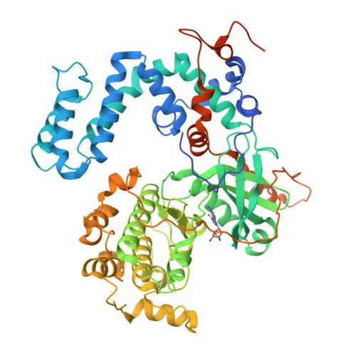
- Encoded by the GRK5 gene (also known as GPRK5), GRK5 is a member of the G protein-coupled receptor kinase (GRK) family that specifically phosphorylates activated GPCRs, initiating desensitization and internalization via beta-arrestin recruitment.
- Beyond GPCRs, GRK5 phosphorylates multiple non-receptor substrates including ST13 (regulating chemokine receptor trafficking), TP53/p53 (inhibiting apoptosis), HDAC5 (promoting nuclear export and enhancing MEF2-mediated transcription), ARRB1, and even LRP6 in Wnt signaling.
- Subcellularly, GRK5 dynamically localizes to the cytoplasm, nucleus, and cell membrane, consistent with its roles in plasma membrane receptor regulation and nuclear transcriptional modulation.
- Tissue distribution is broad but enriched: highest levels are detected in heart, placenta, and lung, with moderate expression in skeletal muscle, brain, liver, pancreas, and kidney.
- The kinase activity is ATP-dependent, and the protein contains lipid-binding domains, enabling membrane association; it is also a phosphoprotein subject to regulatory modifications.
- Functional keywords include apoptosis signaling, Wnt pathway involvement, and serine/threonine kinase activity, underscoring its multifunctional roles in cellular signaling and disease.
Experimental Guidance and Technical Tips
- For Western blotting, prepare whole-cell lysates from tissues with high GRK5 expression (e.g., heart, lung). A band at approximately 68 kDa is expected under reducing conditions; validate with a positive control lysate.
- In immunohistochemistry (IHC-P), standard antigen retrieval (e.g., citrate buffer pH 6.0) may be employed; include known positive tissues such as cardiac muscle and negative controls to assess specificity.
- For immunofluorescence (IF/ICC), cell lines endogenously expressing GRK5 can be used. Fixation/permeabilization conditions should be optimized, and co-localization studies with membrane or nuclear markers may confirm localization dynamics.
- As a polyclonal antibody, batch-to-batch consistency is recommended to be verified for quantitative assays like ELISA. Use recombinant GRK5 protein as a standard.
CamelBio: Your One-Stop Sourcing Bridge
CamelBio supports IVD manufacturers and research labs with a comprehensive portfolio of validated raw materials, including high-quality antibodies, antigen pairs, and ancillary reagents for kinase biology and GPCR signaling research. Whether you require bulk G protein-coupled receptor kinase antibodies, custom recombinant protein production, or assistance in sourcing rare-target raw materials, our expert team ensures seamless integration from concept to clinical application. Partner with CamelBio to accelerate your diagnostic development with reliable and scalable solutions.
REQUEST A QUOTE
Our professional team will reply to you within one business day. Please feel free to contact us!