Monoclonal Antibodies
Anti-GPIX/CD42a/GP9 Rabbit Monoclonal Antibody for WB, IHC-P, ELISA - P14770
Item Number : CM0006582
Price varies based on specs and customizations
- Application
- WB, IHC-P, ELISA
- Cross Reactivity
- Human
- Protein Weight
- 19kDa

Shipping:
Contact us to get shipping details Enjoy On-time Dispatch Guarantee.
Why Choose Us
Easy ordering process, quality products, and dedicated support for your business success.
Core Product Specifications and Parameters
| Parameter | Value |
|---|---|
| Product Name | GPIX/CD42a/GP9 Rabbit mAb |
| Remarks/Alias | GPIX; CD42a; GPIX/CD42a/GP9 |
| Species | Human |
| GeneID | 2815 |
| Immunogen | Recombinant protein of human GPIX/CD42a/GP9 |
| Source | Rabbit |
| Category | Monoclonal Antibodies |
| Application | WB, IHC-P, ELISA |
| Cross Reactivity | Human |
| SWISS | P14770 |
| Protein Weight | 19kDa |
| Shipping | Ice bag |
Biological Background: GPIX/CD42a/GP9 Function and Localization
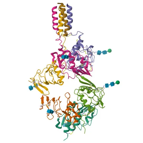
- GPIX, also designated CD42a, is a platelet-specific transmembrane glycoprotein and an essential subunit of the GPIb-V-IX receptor complex, together with GPIbα, GPIbβ, and GPV.
- The GPIb-V-IX complex functions as the high-affinity receptor for von Willebrand factor (vWF), enabling platelet tethering and adhesion to subendothelial collagen at sites of vascular injury under arterial shear conditions.
- This vWF-dependent adhesion is a critical initiating step in primary hemostasis, preventing excessive bleeding.
- GPIX is required for the correct membrane integration and surface expression of the GPIb-IX complex, ensuring proper orientation and functional signaling.
- Loss-of-function mutations in the GP9 gene cause Bernard-Soulier syndrome, an autosomal recessive macrothrombocytopenia with impaired platelet adhesion and bleeding diathesis.
- The protein belongs to the leucine-rich repeat (LRR) superfamily, featuring an N-terminal signal peptide, an extracellular region with LRR motifs, a single-pass transmembrane domain, and a short cytoplasmic tail.
- Post-translational modifications include N-glycosylation and disulfide bond formation, contributing to structural stability and interaction with binding partners.
- Subcellular localization is predominantly at the plasma membrane of platelets and megakaryocytes, with some intracellular distribution likely linked to biosynthesis.
Experimental Guidance and Technical Tips
- This rabbit monoclonal antibody is generated against a recombinant protein encompassing the human GPIX sequence, which may cover the entire extracellular domain; it is suitable for detecting both native and denatured forms.
- For Western blot, the predicted molecular weight is approximately 19 kDa; consider running reducing and non-reducing gels to assess potential homodimerization or complex formation. Note that glycosylation may cause slight band shifts.
- In IHC-P, optimal antigen retrieval conditions (e.g., citrate buffer pH 6.0 or EDTA pH 9.0) should be empirically determined; megakaryocytes and platelets in bone marrow or spleen sections serve as positive controls.
- For ELISA, the antibody can be used as a capture or detection reagent; validate pairing with other antibodies against the GPIb-V-IX complex for sandwich assays.
CamelBio: Your One-Stop Sourcing Bridge
CamelBio serves as a comprehensive procurement partner for IVD manufacturers and researchers, offering validated antibody pairs, monoclonal and polyclonal antibodies, and bulk ancillary reagents to streamline assay development. This anti-GPIX antibody supports hemostasis and platelet biology applications, and CamelBio can facilitate sourcing of related rare targets such as GPIbα, GPV, or custom recombinant proteins to accelerate your diagnostic pipeline from concept to clinic.
Product Datasheet
Anti-GPIX/CD42a/GP9 Rabbit Monoclonal Antibody for WB, IHC-P, ELISA - P14770
REQUEST A QUOTE
Our professional team will reply to you within one business day. Please feel free to contact us!