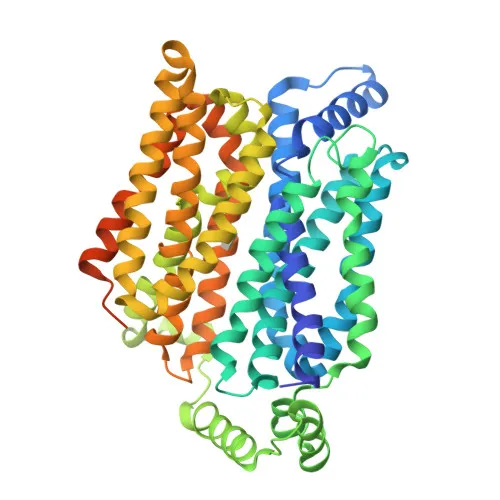
Polyclonal Antibodies
Anti-GLUT1/SLC2A1 Rabbit Polyclonal Antibody for WB, IHC-P, ELISA - P11166
Item Number : CM0026171
Price varies based on specs and customizations
- Application
- WB, IHC-P, ELISA
- Cross Reactivity
- Human, Mouse, Rat
- Protein Weight
- 54kDa

Shipping:
Contact us to get shipping details Enjoy On-time Dispatch Guarantee.
Why Choose Us
Easy ordering process, quality products, and dedicated support for your business success.
Core Product Specifications and Parameters
| Parameter | Details |
|---|---|
| Product Name | GLUT1/SLC2A1 Rabbit pAb |
| Remarks/Alias | CSE; PED; DYT9; GLUT; DYT17; DYT18; EIG12; GLUT1; HTLVR; GLUT-1; SDCHCN; GLUT1DS; GLUT1/SLC2A1 |
| Species | Human |
| GeneID | 6513 |
| Immunogen | Synthetic peptide corresponding to a sequence within amino acids 210-310 of human GLUT1/SLC2A1 (NP_006507.2) |
| Source | Rabbit |
| Category | Polyclonal Antibodies |
| Application | WB, IHC-P, ELISA |
| Cross Reactivity | Human, Mouse, Rat |
| SWISS | P11166 |
| Protein Weight | 54kDa |
| Shipping | Ice bag |
Biological Background: GLUT1/SLC2A1 Function and Localization
- GLUT1/SLC2A1, also known as glucose transporter type 1, erythrocyte/brain GLUT-1, or HepG2 glucose transporter, is encoded by the SLC2A1 gene.
- It functions as a facilitative glucose transporter responsible for constitutive glucose uptake and exhibits broad substrate specificity for aldoses including pentoses and hexoses.
Related references: PMID:10227690 PMID:10954735 PMID:18245775 PMID:19449892 - As the major energy carrier of the brain, GLUT1 ensures facilitative glucose transport across the blood-brain barrier.
Related references: PMID:10227690 - It localizes to the cell membrane, melanosome, and photoreceptor inner segment.
- GLUT1 is detected in erythrocytes at the protein level and expressed at variable levels in many human tissues.
- Post-translational modifications include acetylation and phosphorylation; it is associated with diseases such as dystonia, epilepsy, and hereditary hemolytic anemia.
- In the retina, GLUT1 associates with BSG and NXNL1 to promote cone survival by increasing glucose uptake into photoreceptors.
- It is required for mesendoderm differentiation during development.
Experimental Guidance and Technical Tips
- The immunogen corresponds to a peptide within amino acids 210–310, predicting recognition of an intracellular epitope. For Western blotting, a band around 54 kDa is expected under reducing conditions; include positive controls such as erythrocyte lysates or brain tissue extracts.
- For immunohistochemistry (IHC-P), antigen retrieval may be necessary and membrane staining should be anticipated. Validate staining patterns using known positive tissues.
- In ELISA applications, consider using recombinant GLUT1 protein or peptide as coating antigen and optimize antibody dilution.
- Ensure antibody specificity by including knockout cell lines or siRNA-treated samples where possible.
CamelBio: Your One-Stop Sourcing Bridge
At CamelBio, we provide a comprehensive one-stop sourcing solution for IVD raw materials, offering validated antibody pairs, recombinant proteins, and ancillary reagents for glucose metabolism research. Our portfolio includes high-quality polyclonal antibodies like this anti-GLUT1/SLC2A1 antibody, supporting diagnostic development with reliable and consistent performance.
REQUEST A QUOTE
Our professional team will reply to you within one business day. Please feel free to contact us!