Monoclonal Antibodies
Anti-GFAP Monoclonal Antibody for WB, IF, IHC-P, ELISA - P14136
Item Number : CM0008822
Price varies based on specs and customizations
- Application
- WB, IF/ICC, IF-P, IHC-P, ELISA
- Cross Reactivity
- Mouse, Rat
- Protein Weight
- 50kDa

Shipping:
Contact us to get shipping details Enjoy On-time Dispatch Guarantee.
Why Choose Us
Easy ordering process, quality products, and dedicated support for your business success.
Core Product Specifications and Parameters
| Parameter | Value |
|---|---|
| Product Name | GFAP Rabbit mAb |
| Remarks/Alias | ALXDRD |
| Species | Human |
| GeneID (Human) | 2670 |
| GeneID | 2670 |
| Immunogen | Recombinant fusion protein containing a sequence corresponding to amino acids 60-383 of human GFAP (NP_002046.1) |
| Source | Rabbit |
| Category | Monoclonal Antibodies |
| Application | WB, IF/ICC, IF-P, IHC-P, ELISA |
| Cross Reactivity | Mouse, Rat |
| SWISS | P14136 |
| Protein Weight | 50kDa |
| Shipping | Ice bag |
Biological Background: GFAP Function and Localization
- GFAP (Glial fibrillary acidic protein) is a class-III intermediate filament protein and a well-established marker for astrocytes in the central nervous system.
- It plays a critical role in distinguishing astrocytes from other glial cells during CNS development (UniProt).
- The protein localizes predominantly to the cytoplasm, forming filamentous networks.
- GFAP exhibits tissue-specific expression in cells lacking fibronectin, consistent with its glial cell specificity.
- Post-translational modifications include citrullination, methylation, and phosphorylation, which may regulate filament dynamics and function.
- The GFAP gene undergoes alternative splicing, giving rise to multiple isoforms with potential functional diversity.
- Mutations in GFAP are associated with Alexander disease, a leukodystrophy, highlighting its importance in astrocyte integrity.
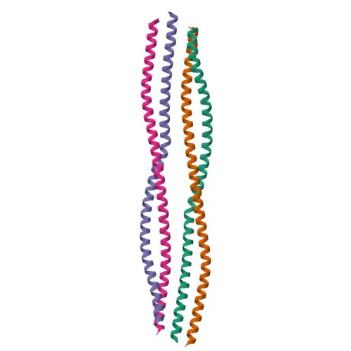
- Keywords from UniProt include: 3D-structure, coiled coil, direct protein sequencing, proteomics identification, reference proteome.
Experimental Guidance and Technical Tips
- The immunogen used to generate this antibody corresponds to amino acids 60-383 of human GFAP, covering a large internal region of the protein. This design may enable recognition of multiple GFAP isoforms and cross-species reactivity, as validated for mouse and rat.
- For Western blotting (WB), the observed band is expected around 50 kDa (theoretical MW ~49.9 kDa). Consider using whole-cell lysates from brain tissue or cultured astrocytes.
- For immunofluorescence (IF/ICC, IF-P) and immunohistochemistry (IHC-P), fixation and permeabilization conditions should be optimized to preserve intermediate filament structure. GFAP staining should produce a typical filamentous cytoplasmic pattern in astrocytes.
- In ELISA applications, this antibody can be used as a capture or detection reagent; pairing with an appropriate matched antibody may improve sensitivity.
- Given the monoclonal nature, batch-to-batch consistency is expected, but validation in the specific experimental setup is advisable.
CamelBio: Your One-Stop Sourcing Bridge
CamelBio offers diagnostic manufacturers, labs, and research institutes streamlined access to IVD raw materials, including validated antibody pairs, bulk monoclonal antibodies, and ancillary reagents for GFAP-based neurological biomarker assays. Whether you are developing GFAP detection kits for brain injury, neurodegeneration, or astrocytic tumor typing, CamelBio can assist with sourcing rare targets and optimizing reagent supply from concept to clinic.
REQUEST A QUOTE
Our professional team will reply to you within one business day. Please feel free to contact us!