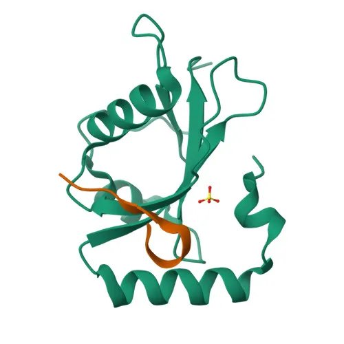
Monoclonal Antibodies
Anti-GABARAPL1 Monoclonal Antibody for WB, IHC-P, ELISA - Q9H0R8
Item Number : CM0005460
Price varies based on specs and customizations
- Application
- WB, IHC-P, ELISA
- Cross Reactivity
- Human, Mouse, Rat
- Protein Weight
- 14kDa/17kDa

Shipping:
Contact us to get shipping details Enjoy On-time Dispatch Guarantee.
Why Choose Us
Easy ordering process, quality products, and dedicated support for your business success.
Core Product Specifications and Parameters
| Parameter | Value |
|---|---|
| Product Name | GABARAPL1 Rabbit mAb |
| Remarks/Alias | GABARAPL1; APG8-LIKE; APG8L; ATG8; ATG8B; ATG8L; GEC1; GABA type A receptor associated protein like 1 |
| Species | Human |
| Gene ID | 23710 |
| Immunogen | Synthetic peptide | A synthetic peptide corresponding to a sequence within amino acids 1-100 of human GABARAPL1. (NP_113600.1). |
| Source | Rabbit |
| Category | Monoclonal Antibodies |
| Application | WB, IHC-P, ELISA |
| Cross Reactivity | Human, Mouse, Rat |
| SWISS | Q9H0R8 |
| Protein Weight | 14kDa/17kDa |
| Shipping | Ice bag |
Biological Background: GABARAPL1 Function and Localization
- GABARAPL1 is a ubiquitin-like modifier that enhances cell-surface expression of kappa-type opioid receptor by promoting its anterograde trafficking. Related references: PMID:16431922
- It is involved in autophagosome formation; while LC3 proteins mediate phagophore elongation, GABARAP subfamily members are critical for later stages of autophagosome maturation. Related references: PMID:20404487
- Through its interaction with the reticulophagy receptor TEX264, GABARAPL1 mediates ER subdomain remodeling into autophagosomes during nutrient stress, facilitating lysosomal degradation for ER turnover. Related references: PMID:31006537 PMID:31006538
- The protein localizes to cytoplasmic vesicles, autophagosome membranes, the cytoskeleton, endoplasmic reticulum, and Golgi apparatus.
- It is ubiquitously expressed, with high levels in brain, heart, liver, kidney, placenta, and skeletal muscle, especially in motoneurons and the substantia nigra pars compacta.
- Alternative splicing produces multiple isoforms; the protein is a lipoprotein with a predicted molecular weight of ~14 kDa, but can be observed at ~17 kDa in tissues.
- Keywords include autophagy, host-virus interaction, proteomics identification, and reference proteome.
Experimental Guidance and Technical Tips
- The immunogen is a synthetic peptide spanning the N-terminal region (amino acids 1-100), which is expected to recognize all splicing isoforms.
- For Western blotting, the predicted molecular weight is ~14 kDa, but due to post-translational modifications, the observed band may be around 17 kDa. Use appropriate gel percentages and loading controls.
- For immunohistochemistry (IHC-P), consider antigen retrieval optimization and validate in known positive tissues (e.g., brain, heart) to confirm specificity.
- The antibody cross-reacts with mouse and rat orthologs, so it can be used in rodent models; however, always validate in the relevant sample system.
- For ELISA, a standard peptide/protein coating buffer and detection system should be applicable; titrate the antibody for optimal signal-to-noise ratio.
CamelBio: Your One-Stop Sourcing Bridge
CamelBio provides diagnostic manufacturers, laboratories, and research institutes with one-stop access to IVD raw materials, from validated antibody pairs and optimized monoclonal/polyclonal antibodies to bulk ancillary reagents and rare target sourcing. This anti-GABARAPL1 monoclonal antibody supports research in autophagy, neuroscience, and cellular trafficking, and CamelBio's integrated supply chain can efficiently meet your project needs at any development stage.
REQUEST A QUOTE
Our professional team will reply to you within one business day. Please feel free to contact us!