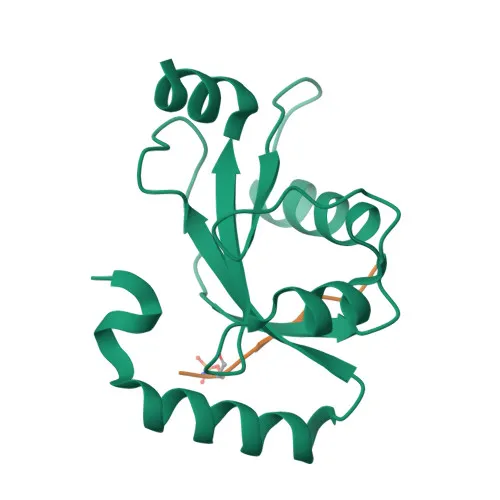
Monoclonal Antibodies
Anti-FUNDC1 Rabbit Monoclonal Antibody for WB, IP, IHC-P, ELISA - Q8IVP5
Item Number : CM0011310
Price varies based on specs and customizations
- Application
- WB, IP, IHC-P, ELISA
- Cross Reactivity
- Human, Mouse, Rat
- Protein Weight
- 17kDa

Shipping:
Contact us to get shipping details Enjoy On-time Dispatch Guarantee.
Why Choose Us
Easy ordering process, quality products, and dedicated support for your business success.
Core Product Specifications and Parameters
| Parameter | Value |
|---|---|
| Product Name | FUNDC1 Rabbit mAb |
| Remarks/Alias | Fundc1 |
| Species | Human |
| GeneID | 139341 |
| Immunogen | Synthetic peptide corresponding to a sequence within amino acids 1-100 of human FUNDC1 (NP_776155.1) |
| Source | Rabbit |
| Category | Monoclonal Antibodies |
| Application | WB, IP, IHC-P, ELISA |
| Cross Reactivity | Human, Mouse, Rat |
| SWISS | Q8IVP5 |
| Protein Weight | 17kDa |
| Shipping | Ice bag |
Biological Background: FUNDC1 Function and Localization
- FUNDC1 is an integral mitochondrial outer-membrane protein that mediates the formation of mitochondria-associated endoplasmic reticulum membranes (MAMs), facilitating inter-organelle communication. Related references: PMID:33972548
- Through MAM formation, FUNDC1 regulates intracellular Ca2+ signaling and vascular endothelial growth factor receptor KDR/VEGFR2 expression, thereby promoting angiogenesis and neoangiogenesis. Related references: PMID:33972548
- FUNDC1 functions as an activator of hypoxia-induced mitophagy by interacting with and recruiting LC3 protein family members to mitochondria, ensuring mitochondrial quality control. Related references: PMID:22267086 PMID:24671035 PMID:24746696
- During hypoxia, FUNDC1 recruits DRP1 to ER-mitochondria contact sites, promoting DRP1 oligomerization and GTPase activity, which drives mitochondrial fission. Related references: PMID:27145933 PMID:33978709
- FUNDC1 contributes to hepatic ferroptosis by interacting with glutathione peroxidase GPX4, facilitating its mitochondrial import via the TOM/TIM complex and subsequent degradation through mitophagy. Related references: PMID:36828120
- The full-length human FUNDC1 protein has a predicted molecular weight of approximately 17 kDa and localizes to the mitochondrial outer membrane. It is widely expressed across tissues.
- Keywords: autophagy, isopeptide bond, membrane, mitochondrion outer membrane, phosphoprotein, reference proteome, transmembrane helix, Ubl conjugation.
Experimental Guidance and Technical Tips
- The immunogen corresponds to the N-terminal region (amino acids 1-100) of human FUNDC1; verify sequence conservation in mouse and rat orthologs if cross-reactivity is critical.
- For western blot, a band of approximately 17 kDa is expected; consider using mitochondrial-enriched fractions or appropriate positive controls (e.g., HeLa, HEK293, or mouse liver lysates).
- For immunohistochemistry on paraffin sections, antigen retrieval may be required; validate staining specificity in tissues with known FUNDC1 expression.
- For immunoprecipitation, pilot experiments should optimize antibody-to-lysate ratio and incubation conditions.
- The rabbit monoclonal format ensures high specificity and batch-to-batch consistency; confirm optimal working dilutions for each application in your experimental system.
CamelBio: Your One-Stop Sourcing Bridge
CamelBio supports your mitophagy and angiogenesis research with validated antibody reagents like this FUNDC1 rabbit monoclonal antibody, along with a comprehensive portfolio of IVD raw materials, including antibody pairs, ancillary reagents, and rare-target sourcing. From early-stage development to clinical application, our integrated supply chain and technical expertise help streamline your diagnostic assay creation.
Product Datasheet
Anti-FUNDC1 Rabbit Monoclonal Antibody for WB, IP, IHC-P, ELISA - Q8IVP5
REQUEST A QUOTE
Our professional team will reply to you within one business day. Please feel free to contact us!