Polyclonal Antibodies
Anti-FUNDC1 Polyclonal Antibody for WB, IHC-P, ELISA - Q8IVP5
Item Number : CM0019396
Price varies based on specs and customizations
- Application
- WB, IHC-P, ELISA
- Cross Reactivity
- Human, Mouse
- Protein Weight
- 17kDa

Shipping:
Contact us to get shipping details Enjoy On-time Dispatch Guarantee.
Why Choose Us
Easy ordering process, quality products, and dedicated support for your business success.
Core Product Specifications and Parameters
| Parameter | Value |
|---|---|
| Product Name | FUNDC1 Rabbit pAb |
| Remarks/Alias | FUNDC1 |
| Species | Human |
| GeneID (human) | 139341 |
| GeneID | 139341 |
| Immunogen | Synthetic Peptide - A synthetic peptide corresponding to a sequence within amino acids 1-155 of human FUNDC1 (NP_776155.1). |
| Source | Rabbit |
| Category | Polyclonal Antibodies |
| Application | WB, IHC-P, ELISA |
| Cross Reactivity | Human, Mouse |
| SWISS | Q8IVP5 |
| Protein Weight | 17kDa |
| Shipping | Ice bag |
Biological Background: FUNDC1 Function and Localization
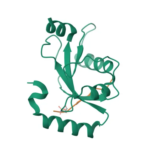
- FUNDC1 (FUN14 domain-containing protein 1) is a mitochondrial outer membrane protein that integrates autophagy and mitochondrial quality control.
- It mediates formation of mitochondria-associated ER membranes (MAMs) and promotes angiogenesis through regulation of intracellular Ca(2+) and VEGFR2 expression. Related references: PMID:33972548
- As a mitophagy receptor, FUNDC1 recruits LC3 family proteins to mitochondria during hypoxia, facilitating selective autophagic degradation. Related references: PMID:22267086 | PMID:24671035 | PMID:24746696
- It also interacts with DRP1 at ER-mitochondria contact sites, driving mitochondrial fission under hypoxic conditions. Related references: PMID:27145933 | PMID:33978709
- Recent evidence shows FUNDC1 is involved in hepatic ferroptosis by binding to GPX4 and targeting it for mitophagic degradation. Related references: PMID:36828120
- Subcellularly, FUNDC1 is anchored in the mitochondrial outer membrane and is widely expressed in tissues.
- Post-translational modifications include phosphorylation and ubiquitin conjugation, as indicated by UniProt keywords.
Experimental Guidance and Technical Tips
- The immunogen is a synthetic peptide corresponding to the N-terminal region (aa 1-155) of human FUNDC1; this sequence is expected to detect full-length FUNDC1 and potentially any N-terminal isoforms.
- For Western blotting, the predicted molecular weight is 17 kDa; consider using reducing conditions and validate band detection in relevant human or mouse cell/tissue lysates.
- In IHC-P, antigen retrieval (e.g., citrate buffer pH 6.0) may be required; include appropriate positive and negative tissue controls.
- For ELISA, the antibody can be used for direct or indirect assays; titrate the antibody concentration for optimal signal-to-noise ratio.
CamelBio: Your One-Stop Sourcing Bridge
CamelBio serves as a one-stop sourcing bridge for IVD raw materials, offering validated antibodies, bulk ancillary reagents, and rare target raw-material procurement. For researchers focused on mitochondrial biology and autophagy, we provide reliable polyclonal antibodies like this anti-FUNDC1 antibody to support your assay development and biomarker discovery workflows. Contact us to explore customized sourcing solutions tailored to your pipeline.
REQUEST A QUOTE
Our professional team will reply to you within one business day. Please feel free to contact us!