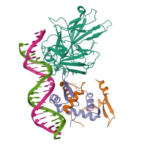
Monoclonal Antibodies
Anti-FOXP3 Monoclonal Antibody for WB, IP, ELISA - Q9BZS1
Item Number : CM0005107
Price varies based on specs and customizations
- Application
- WB, IP, ELISA
- Cross Reactivity
- Human
- Protein Weight
- 47kDa

Shipping:
Contact us to get shipping details Enjoy On-time Dispatch Guarantee.
Why Choose Us
Easy ordering process, quality products, and dedicated support for your business success.
Core Product Specifications and Parameters
| Parameter | Value |
|---|---|
| Product Name | FOXP3 Rabbit mAb |
| Remarks/Alias | JM2; AIID; IPEX; PIDX; XPID; DIETER; FOXP3 |
| Species | Human |
| GeneID (Human) | 50943 |
| GeneID | 50943 |
| Immunogen | Recombinant protein |
| Source | Rabbit |
| Category | Monoclonal Antibodies |
| Application | WB, IP, ELISA |
| Cross Reactivity | Human |
| SWISS | Q9BZS1 |
| Protein Weight | 47kDa |
| Shipping | Ice bag |
Biological Background: FOXP3 Function and Localization
- Forkhead box protein P3 (FOXP3), also known as Scurfin, is a transcription factor essential for immune regulation.
- Crucial for the development and inhibitory function of regulatory T-cells (Treg) and for maintaining immune homeostasis by ensuring Treg suppressive function and stability, and modulating conventional T-cell expansion and function. Related references: PMID:17377532 PMID:21458306 PMID:23947341 PMID:23169781
- Functions as a transcriptional repressor or activator, depending on interactions with cofactors such as histone acetylases and deacetylases. Related references: PMID:17377532 PMID:21458306 PMID:23947341
- Coordinates expression of immune-related genes: activates CTLA4 and TNFRSF18 while repressing IL2 and IFNG. Related references: PMID:17377532 PMID:21458306 PMID:23947341
- Suppresses cytokine production by inhibiting RELA and NFATC2, and mediates IL2 repression via interactions with KAT5 and HDAC7. Related references: PMID:15790681 PMID:17360565
- Interacts with RUNX1 to regulate target genes, and with IKZF4 to repress IL2 and IFNG. Related references: PMID:17377532
- Inhibits Th17 differentiation by antagonizing RORC, favoring Treg development. Related references: PMID:18368049
- Subcellular localization: Predominantly nuclear, also found in cytoplasm. Subject to post-translational modifications including acetylation and phosphorylation, which modulate its activity.
Experimental Guidance and Technical Tips
- The immunogen corresponds to the C-terminal region (aa 286-431) of human FOXP3, which includes the forkhead DNA-binding domain. This region is highly conserved; however, cross-reactivity data is currently limited to human samples.
- For western blotting, the predicted molecular weight of FOXP3 is approximately 47 kDa. A nuclear-rich lysate from Treg cells is recommended for clear detection.
- For immunoprecipitation, consider using nuclear extraction protocols to enrich for transcription factors and validate the antibody’s binding capacity in the relevant cellular context.
- In ELISA, using immobilized recombinant FOXP3 protein or lysates from FOXP3-expressing cells can help assess binding. Titration of the detection antibody and appropriate blocking are advised.
- Given the predominantly nuclear localization, nuclear fractionation may enhance signal in both WB and IP applications.
CamelBio: Your One-Stop Sourcing Bridge
CamelBio connects diagnostic manufacturers, labs, and research institutes with a comprehensive portfolio of IVD raw materials, technical services, and consulting—from concept to clinic. This FOXP3 Rabbit mAb supports immunology research and Treg-related assay development, reflecting our commitment to supplying validated antibody pairs, bulk ancillary reagents, and rare-target raw materials. Whatever your sourcing needs, including optimized monoclonal/polyclonal antibodies for diagnostic pipelines, CamelBio streamlines procurement to accelerate your project timeline.
REQUEST A QUOTE
Our professional team will reply to you within one business day. Please feel free to contact us!