Polyclonal Antibodies
Anti-Folate Binding Protein (FOLR1) Rabbit Polyclonal Antibody for WB - P15328
Item Number : CM0023521
Price varies based on specs and customizations
- Application
- WB, IHC-P, IF/ICC, ELISA
- Cross Reactivity
- Human, Mouse, Rat
- Protein Weight
- 30kDa

Shipping:
Contact us to get shipping details Enjoy On-time Dispatch Guarantee.
Why Choose Us
Easy ordering process, quality products, and dedicated support for your business success.
Core Product Specifications and Parameters
| Parameter | Value |
|---|---|
| Product Name | Folate Binding Protein(FBP) / FOLR1 Rabbit pAb |
| Remarks/Alias | FBP; FOLR; NCFTD; FRalpha; Folate Binding Protein(FBP) / FOLR1 |
| Species | Human |
| Gene ID (Human) | 2348 |
| Gene ID | 2348 |
| Immunogen | Synthetic peptide |
| Source | Rabbit |
| Category | Polyclonal Antibodies |
| Application | WB, IHC-P, IF/ICC, ELISA |
| Cross Reactivity | Human, Mouse, Rat |
| SWISS | P15328 |
| Protein Weight | 30kDa |
| Shipping | Ice bag |
Biological Background: Folate Receptor Alpha (FOLR1) Function and Localization
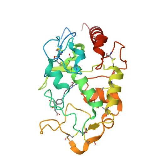
- FOLR1 encodes folate receptor alpha (FRα), also known as adult folate-binding protein, ovarian tumor-associated antigen MOv18, and KB cells FBP.
- FRα is a high-affinity folate receptor that mediates cellular uptake of 5-methyltetrahydrofolate and folate analogs via receptor-mediated endocytosis. Related references: PMID:19074442, PMID:23851396, PMID:23934049, PMID:2527252
- Following endocytosis, acidic pH in the endosome triggers a conformational change that reduces folate binding affinity, leading to folate release into the cytoplasm. Related references: PMID:8567728
- FRα is a GPI-anchored glycoprotein and lipoprotein, as indicated by its keywords and structural features.
- Subcellularly, FRα localizes to the cell membrane (apical and basolateral surfaces), cytoplasmic vesicles, clathrin-coated vesicles, and endosomes. A secreted form has also been reported.
- FRα is primarily expressed in epithelial tissues including kidney, lung, and cerebellum, with increased expression in malignant tissues; it is also detected in placenta and thymus epithelium.
- The protein contains disulfide bonds and undergoes glycosylation, essential for its stability and membrane targeting.
- FRα is required for normal embryonic development and cell proliferation (By similarity).
Experimental Guidance and Technical Tips
- The immunogen corresponds to the N-terminal region (aa 1-100) of human FOLR1, which includes extracellular domains; thus the antibody may target surface-exposed epitopes, making it suitable for IHC-P and IF/ICC protocols, but optimization of permeabilization may be needed for intracellular detection.
- For Western blot, a band of approximately 30 kDa is expected; however, due to glycosylation and other post-translational modifications, the observed molecular weight may vary, and validation in the relevant species or tissue lysates is recommended.
- In ELISA applications, consider using recombinant FOLR1 protein or known folate-binding protein standards as positive controls.
- Cross-reactivity with mouse and rat has been reported, but sequence alignment of the immunogen region should be confirmed if using in these species, and antibody titration is advised.
CamelBio: Your One-Stop Sourcing Bridge
As your integrated sourcing partner, CamelBio streamlines procurement of high-quality antibodies like this anti-FOLR1 polyclonal antibody, along with validated antibody pairs, recombinant proteins, and ancillary reagents for folate metabolism and oncology diagnostics. We offer bulk orders, customized panel development, and technical consultation to accelerate your assay development from research to commercial scale.
Product Datasheet
Anti-Folate Binding Protein (FOLR1) Rabbit Polyclonal Antibody for WB - P15328
REQUEST A QUOTE
Our professional team will reply to you within one business day. Please feel free to contact us!