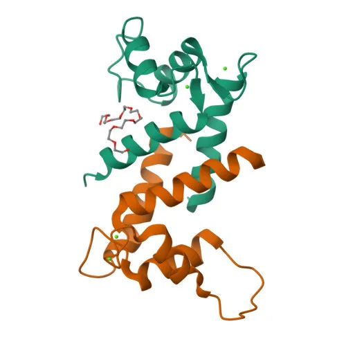
Polyclonal Antibodies
Anti-FLG Rabbit Polyclonal Antibody for WB, ELISA - P20930
Item Number : CM0022028
Price varies based on specs and customizations
- Application
- WB, ELISA
- Cross Reactivity
- Human, Mouse, Rat
- Protein Weight
- 435 kDa

Shipping:
Contact us to get shipping details Enjoy On-time Dispatch Guarantee.
Why Choose Us
Easy ordering process, quality products, and dedicated support for your business success.
Core Product Specifications and Parameters
| Parameter | Value |
|---|---|
| Product Name | FLG Rabbit Polyclonal Antibody |
| Remarks / Alias | FLG1; ATOD2; FLG-1; FLG |
| Species | Human |
| Gene ID (Human) | 2312 |
| Gene ID | 2312 |
| Immunogen | Recombinant fusion protein containing a sequence corresponding to amino acids 1-92 of human FLG (NP_002007.1). |
| Source | Rabbit |
| Category | Polyclonal Antibodies |
| Application | WB, ELISA |
| Cross Reactivity | Human, Mouse, Rat |
| SWISS | P20930 |
| Protein Weight | 435 kDa |
| Shipping | Ice bag |
Biological Background: Filaggrin Function and Localization
- Filaggrin (FLG) is a large, histidine-rich, basic protein that aggregates keratin intermediate filaments in the epidermis, forming the structural matrix of the cornified cell envelope.
- It promotes disulfide-bond formation among intermediate filaments, contributing to the mechanical strength and barrier function of the skin.
- FLG is expressed predominantly in the stratum granulosum of the epidermis, where it is synthesized as a giant precursor, profilaggrin, which is subsequently proteolytically processed into active filaggrin monomers.
- Subcellularly, filaggrin localizes to cytoplasmic keratohyalin granules in granular layer keratinocytes.
- The protein contains multiple filaggrin repeats that facilitate keratin binding and filament aggregation, and its function is regulated by calcium ions and post-translational modifications like citrullination.
- As a developmental protein, FLG plays a critical role in the terminal differentiation of keratinocytes and the formation of a waterproof skin barrier.
- Mutations in the FLG gene are a major predisposing factor for ichthyosis vulgaris and atopic dermatitis, highlighting its clinical significance in skin disorders.
Experimental Guidance and Technical Tips
- The immunogen corresponds to amino acids 1-92 of human FLG (N-terminal region). This region may be conserved across species, consistent with reported cross-reactivity with mouse and rat. Validate cross-reactivity in your sample system if using non-human models.
- Recommended applications include Western blotting (WB) and ELISA. For WB, note the large molecular weight (~435 kDa); consider using a low-percentage gel and extended transfer times to ensure efficient protein transfer.
- Due to the high molecular weight and repetitive nature of filaggrin, consider using fresh or well-preserved samples to avoid protein degradation, and include protease inhibitors during sample preparation.
CamelBio: Your One-Stop Sourcing Bridge
CamelBio serves as a comprehensive sourcing partner for diagnostic manufacturers and research laboratories, offering validated antibody pairs, optimized monoclonal and polyclonal antibodies, and bulk ancillary reagents. This anti-FLG antibody supports research in epidermal differentiation, skin barrier function, and dermatological diseases. For specialty targets like filaggrin, we provide custom antibody development and rare target raw-material sourcing to accelerate your assay development from concept to clinic.
REQUEST A QUOTE
Our professional team will reply to you within one business day. Please feel free to contact us!