Polyclonal Antibodies
Anti-FIG4 Polyclonal Antibody for WB, ELISA - Q92562
Item Number : CM0013176
Price varies based on specs and customizations
- Application
- WB, ELISA
- Cross Reactivity
- Human, Mouse
- Protein Weight
- 104kDa

Shipping:
Contact us to get shipping details Enjoy On-time Dispatch Guarantee.
Why Choose Us
Easy ordering process, quality products, and dedicated support for your business success.
Core Product Specifications and Parameters
| Parameter | Value |
|---|---|
| Product Name | FIG4 Rabbit pAb |
| Remarks/Alias | BOP; YVS; BTOP; SAC3; ALS11; CMT4J; KIAA0274; dJ249I4.1; FIG4 |
| Species | Human |
| Gene ID (Human) | 9896 |
| Immunogen | Recombinant fusion protein containing a sequence corresponding to amino acids 608-907 of human FIG4 (NP_055660.1) |
| Source | Rabbit |
| Category | Polyclonal Antibodies |
| Application | WB, ELISA |
| Cross Reactivity | Human, Mouse |
| SWISS | Q92562 |
| Protein Weight | 104kDa |
| Shipping | Ice bag |
Biological Background: FIG4 Function and Localization
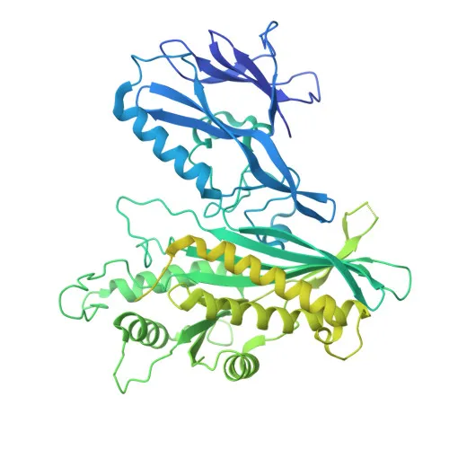
- FIG4, also known as SAC3 or KIAA0274, is a polyphosphoinositide phosphatase and a core component of the PI(3,5)P2 regulatory complex, crucial for phosphatidylinositol metabolism and endolysosomal signaling.
- It functions as a dual specificity phosphatase: it catalyzes the dephosphorylation of PtdIns(3,5)P2 to generate PtdIns(3)P, and also acts as a serine-protein phosphatase on PIKfyve to stimulate its lipid kinase activity, thereby fine-tuning PtdIns(3,5)P2 levels. Related references: PMID:17556371 PMID:33098764
- The protein localizes predominantly to the endosome membrane, consistent with its role in regulating endosomal phosphoinositide pools.
- Mutations in the FIG4 gene are linked to severe neurodegenerative diseases, including amyotrophic lateral sclerosis type 11 (ALS11) and Charcot-Marie-Tooth disease type 4J (CMT4J), making it a clinically significant target.
- In vitro, FIG4 can also hydrolyze other D5-phosphorylated polyphosphoinositides such as PtdIns(3,4,5)P3 and PtdIns(4,5)P2, albeit with lower preference compared to PtdIns(3,5)P2. Related references: PMID:17556371
- The molecular weight of human FIG4 is approximately 104 kDa (103,635 Da based on sequence), aligning with the protein's complex domain architecture.
Experimental Guidance and Technical Tips
- For Western blot, use whole cell lysates from human or mouse samples (e.g., HeLa, NIH/3T3) due to the antibody's confirmed cross-reactivity with these species.
- Titrate the antibody concentration for each application; a typical starting dilution range for polyclonal antibodies is 1:500–1:2000 for WB and 1:1000–1:5000 for indirect ELISA, but optimal conditions must be empirically determined.
- The immunogen spans the C-terminal domain (aa 608–907), so the antibody should detect full-length FIG4 along with relevant isoforms.
- Because FIG4 is an endosome-associated protein, subcellular fractionation or membrane protein enrichment may improve detection sensitivity in WB.
CamelBio: Your One-Stop Sourcing Bridge
CamelBio acts as a comprehensive supplier of IVD raw materials, providing validated antibodies, optimized polyclonal and monoclonal reagents, and bulk ancillary products to accelerate your assay development. This anti-FIG4 antibody supports research into neurodegeneration and phosphoinositide signaling, and we can assist with sourcing complementary reagents such as antigen controls and detection systems. From concept to clinic, CamelBio offers integrated technical consulting and one-stop procurement to streamline your diagnostic pipeline.
REQUEST A QUOTE
Our professional team will reply to you within one business day. Please feel free to contact us!