Monoclonal Antibodies
Anti-FGFR4 Monoclonal Antibody for WB, ELISA - P22455
Item Number : CM0004201
Price varies based on specs and customizations
- Application
- WB, ELISA
- Cross Reactivity
- Human, Mouse, Rat
- Protein Weight
- 88 kDa

Shipping:
Contact us to get shipping details Enjoy On-time Dispatch Guarantee.
Why Choose Us
Easy ordering process, quality products, and dedicated support for your business success.
Easy Process
Quality Assured
Dedicated Support
Core Product Specifications and Parameters
| Parameter | Value |
|---|---|
| Product Name | FGFR4 Rabbit mAb |
| Remarks/Alias | TKF, JTK2, CD334, FGFR4 |
| Species | Human |
| GeneID (human) | 2264 |
| GeneID | 2264 |
| Immunogen | Synthetic peptide (aa 700-802 of human FGFR4, P22455) |
| Source | Rabbit |
| Category | Monoclonal Antibodies |
| Application | WB, ELISA |
| Cross Reactivity | Human, Mouse, Rat |
| SWISS | P22455 |
| Protein Weight | 88 kDa |
| Shipping | Ice bag |
Biological Background: FGFR4 Function and Localization
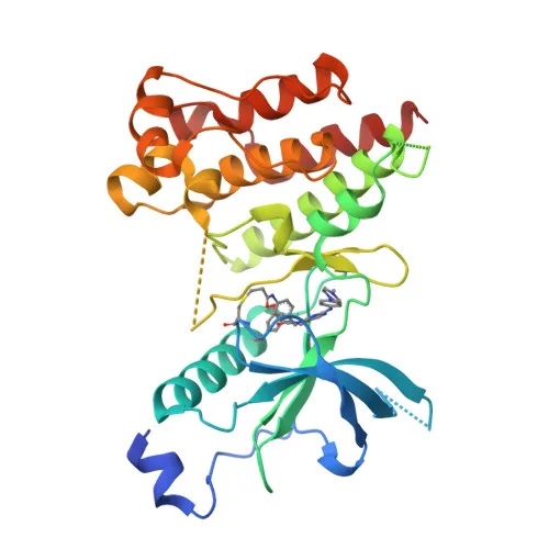
- Fibroblast growth factor receptor 4 (FGFR4) is a tyrosine-protein kinase that serves as a cell-surface receptor for fibroblast growth factors (FGFs). It regulates cell proliferation, differentiation, migration, lipid metabolism, bile acid biosynthesis, glucose uptake, vitamin D metabolism, and phosphate homeostasis.
- Ligand binding triggers activation of downstream signaling cascades, including PLCG1 and FRS2 pathways, leading to production of second messengers (DAG, IP3) and activation of RAS-MAPK and AKT pathways.
- FGFR4 signaling is downregulated by receptor internalization and degradation; the matrix protease MMP14 promotes internalization and lysosomal degradation of FGFR4. Constitutively activating mutations or impaired inactivation lead to aberrant signaling.
- The receptor localizes to the cell membrane, endosomes, endoplasmic reticulum, and a secreted form exists. It is expressed in gastrointestinal epithelial cells, pancreas, and gastric and pancreatic cancer cell lines.
- Alternative splicing produces multiple isoforms, and the protein undergoes post-translational modifications including glycosylation, phosphorylation, and ubiquitin conjugation (Ubl conjugation).
- Keywords: ATP-binding, kinase, transmembrane helix, immunoglobulin domain, disulfide bond, glycoprotein, phosphoprotein, and receptor.
Experimental Guidance and Technical Tips
- Given the immunogen is a synthetic peptide corresponding to the C‑terminal region (amino acids 700–802), the antibody is likely to recognize full‑length FGFR4 as well as isoforms containing this epitope. Consider sequence alignment to verify epitope conservation across species.
- For Western blot, a band at approximately 88 kDa is expected for FGFR4 in human, mouse, and rat samples. Use appropriate gel percentage (e.g., 8–10% SDS-PAGE) for optimal resolution. Consider starting with a primary antibody dilution range of 1:1,000–1:5,000 and validate in your experimental system.
- For ELISA, titrate the antibody to determine the optimal coating concentration and detection conditions. Ensure recombinant FGFR4 protein or peptide antigen matches the immunogen region to avoid false negatives.
CamelBio: Your One-Stop Sourcing Bridge
- CamelBio partners with diagnostic manufacturers, laboratories, and research institutes to provide a seamless supply of IVD raw materials, technical services, and consultative support. This anti‑FGFR4 monoclonal antibody is ideally suited for studies in cancer, metabolism, and cell signaling.
- Beyond individual antibodies, CamelBio offers validated antibody pairs, optimized monoclonal and polyclonal antibodies, bulk ancillary reagents, and customized sourcing for rare or difficult‑to‑find targets, helping you streamline your assay development from concept to clinic.
REQUEST A QUOTE
Our professional team will reply to you within one business day. Please feel free to contact us!