Monoclonal Antibodies
Anti-FGF1 Monoclonal Antibody for WB, IF/ICC, ELISA - P05230
Item Number : CM0008800
Price varies based on specs and customizations
- Application
- WB, IF/ICC, ELISA
- Cross Reactivity
- Human, Mouse, Rat
- Protein Weight
- 17kDa

Shipping:
Contact us to get shipping details Enjoy On-time Dispatch Guarantee.
Why Choose Us
Easy ordering process, quality products, and dedicated support for your business success.
Core Product Specifications and Parameters
| Parameter | Value |
|---|---|
| Product Name | FGF1 Rabbit mAb |
| Remarks/Alias | AFGF; ECGF; FGFA; ECGFA; ECGFB; FGF-1; HBGF1; HBGF-1; GLIO703; ECGF-beta; FGF-alpha; FGF1 |
| Species | Human |
| GeneID (Human) | 2246 |
| GeneID | 2246 |
| Immunogen | Synthetic Peptide (A synthetic peptide corresponding to a sequence within amino acids 56-155 of human FGF1 (P05230)) |
| Source | Rabbit |
| Category | Monoclonal Antibodies |
| Application | WB, IF/ICC, ELISA |
| Cross Reactivity | Human, Mouse, Rat |
| SWISS | P05230 |
| Protein Weight | 17kDa |
| Shipping | Ice bag |
Biological Background: FGF1 Function and Localization
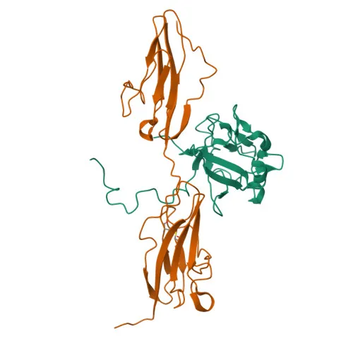
- FGF1, also known as acidic fibroblast growth factor (aFGF) or heparin-binding growth factor 1 (HBGF-1), is a member of the fibroblast growth factor family and functions as a potent mitogen involved in cell survival, proliferation, differentiation, migration, and angiogenesis.
- Through binding to FGFR1 and integrin ITGAV:ITGB3, FGF1 triggers receptor dimerization and autophosphorylation, subsequently activating downstream signaling cascades such as MAPK/ERK and AKT pathways.
Related references: PMID:18441324, PMID:20422052 - FGF1 plays a direct role in promoting angiogenesis, a critical process in development and tissue repair.
Related references: PMID:23469107 - The protein is localized in multiple cellular compartments including the cytoplasm, nucleus, and is also secreted extracellularly.
- Expression is highest in the kidney and brain, with lower levels detected in heart and skeletal muscle.
- Key structural features include heparin-binding domains, and the protein undergoes post-translational modifications such as acetylation and phosphorylation. Multiple isoforms exist due to alternative splicing.
- It is involved in developmental processes, which aligns with its classification as a developmental protein.
Experimental Guidance and Technical Tips
- For Western blot (WB), expect a band at approximately 17 kDa. Use reducing conditions and appropriate positive controls from human, mouse, or rat cell lysates.
- In immunofluorescence (IF/ICC), validate the staining pattern against the known subcellular distribution (cytoplasmic and nuclear). Heparin co-incubation may influence localization due to FGF1's heparin-binding property.
- For ELISA applications, optimize coating and detection conditions empirically; the antibody may function as a capture or detection reagent.
- The immunogen peptide (aa 56-155) covers a region conserved across human, mouse, and rat, supporting the observed cross-reactivity. Validate performance in your specific model system.
- Since FGF1 is a secreted heparin-binding protein, consider heparin-affinity enrichment when preparing samples from tissue extracts or cell supernatants.
- Upon receipt, store the antibody as recommended (shipped on ice bags), typically at -20°C.
CamelBio: Your One-Stop Sourcing Bridge
CamelBio supports diagnostic manufacturers and research laboratories by providing a comprehensive range of IVD raw materials and technical services from concept to clinic. For FGF1-targeted projects, including cell signaling and angiogenesis studies, CamelBio offers access to validated antibodies, recombinant proteins, and ancillary reagents. We help streamline your sourcing needs for monoclonal antibodies, bulk reagents, and customized solutions.
REQUEST A QUOTE
Our professional team will reply to you within one business day. Please feel free to contact us!