Monoclonal Antibodies
Anti-Eph receptor B3 (EPHB3) Monoclonal Antibody for WB, ELISA - P54753
Item Number : CM0003021
Price varies based on specs and customizations
- Application
- WB, ELISA
- Cross Reactivity
- Human, Mouse, Rat
- Protein Weight
- 110kDa

Shipping:
Contact us to get shipping details Enjoy On-time Dispatch Guarantee.
Why Choose Us
Easy ordering process, quality products, and dedicated support for your business success.
Core Product Specifications and Parameters
| Parameter | Value |
|---|---|
| Product Name | Eph receptor B3 (EPHB3) Rabbit mAb |
| Remarks/Alias | EK2; ETK2; HEK2; TYRO6; Eph receptor B3 (EPHB3) |
| Species | Human |
| GeneID | 2049 (Human) |
| Immunogen | Synthetic peptide corresponding to a sequence within amino acids 50-150 of human Eph receptor B3 (EPHB3) (P54753) |
| Source | Rabbit |
| Category | Monoclonal Antibodies |
| Application | WB, ELISA |
| Cross Reactivity | Human, Mouse, Rat |
| SWISS | P54753 |
| Protein Weight | 110kDa |
| Shipping | Ice bag |
Biological Background: Eph receptor B3 (EPHB3) Function and Localization
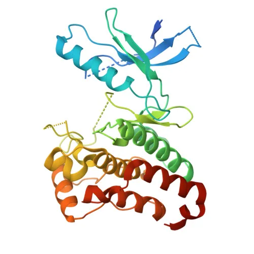
- Eph receptor B3 (EPHB3), also known as EK2, ETK2, HEK2, and TYRO6, is a member of the ephrin receptor subfamily of receptor tyrosine kinases.
- EPHB3 binds promiscuously to transmembrane ephrin-B ligands on adjacent cells, mediating contact-dependent bidirectional signaling: forward signaling through the receptor and reverse signaling through the ephrin.
- It functions redundantly with EPHB2 in axon guidance during development, regulating the formation of major interhemispheric connections such as the corpus callosum and anterior commissure.
- EPHB3 also contributes to dendritic spine development, excitatory synapse formation, and cell migration and positioning in processes including angiogenesis, palate formation, and thymic epithelium development.
- Forward and reverse signaling through the EFNB2/EPHB3 complex regulate cell adhesion and migration during urethra tubularization and cloaca septation.
- In the intestinal epithelium, EPHB3 helps segregate progenitor cells from differentiated cells within the crypt.
- Subcellularly, EPHB3 localizes to the cell membrane and dendrites.
- As a kinase, it contains characteristic features such as an ATP-binding domain, and is modified by glycosylation, phosphorylation, and ubiquitination.
Experimental Guidance and Technical Tips
- The immunogen is a synthetic peptide corresponding to amino acids 50–150 within the N-terminal extracellular region of human EPHB3. This region is likely exposed and may be suitable for detecting both full-length and soluble forms of the receptor, but cross-reactivity with other ephrin receptors should be validated.
- For Western blot, the predicted molecular weight is 110 kDa; consider optimizing gel percentage, transfer conditions, and blocking buffers to resolve this relatively large protein.
- In ELISA, the antibody may be used for detection of recombinant or endogenous EPHB3; titrate appropriately and include relevant controls for background.
- Since the antibody shows cross-reactivity with mouse and rat, it can be applied in model organisms, but confirm reactivity in your specific tissue or cell lysates due to potential species-specific variations.
- Given EPHB3's role in development and cancer, consider testing in tissue samples or cell lines where EPHB3 expression is expected (e.g., brain, intestinal crypts, certain tumor cells).
CamelBio: Your One-Stop Sourcing Bridge
CamelBio provides diagnostic manufacturers, labs, and research institutes with one-stop access to IVD raw materials, technical services, and consulting—covering every stage from concept to clinic. Our portfolio includes validated monoclonal antibodies like this anti-EPHB3 clone, ideal for neurological and developmental research applications. We also supply bulk ancillary reagents and can assist in sourcing rare-target raw materials to accelerate your assay development.
Product Datasheet
Anti-Eph receptor B3 (EPHB3) Monoclonal Antibody for WB, ELISA - P54753
REQUEST A QUOTE
Our professional team will reply to you within one business day. Please feel free to contact us!