Polyclonal Antibodies
Anti-EpCAM Rabbit Polyclonal Antibody for WB, IF/ICC, ELISA - P16422
Item Number : CM0014628
Price varies based on specs and customizations
- Application
- WB, IF/ICC, ELISA
- Cross Reactivity
- Human, Mouse, Rat
- Protein Weight
- 35kDa

Shipping:
Contact us to get shipping details Enjoy On-time Dispatch Guarantee.
Why Choose Us
Easy ordering process, quality products, and dedicated support for your business success.
Core Product Specifications and Parameters
| Parameter | Value |
|---|---|
| Product Name | EpCAM Rabbit pAb |
| Remarks/Alias | ESA; KSA; M4S1; MK-1; DIAR5; EGP-2; EGP40; KS1/4; MIC18; TROP1; BerEp4; EGP314; HNPCC8; LYNCH8; MOC-31; Ber-Ep4; TACSTD1; EpCAM |
| Species | Human |
| GeneID (Human) | 4072 |
| GeneID | 4072 |
| Immunogen | Recombinant fusion protein containing a sequence corresponding to amino acids 24-265 of human EpCAM (NP_002345.2) |
| Source | Rabbit |
| Category | Polyclonal Antibodies |
| Application | WB, IF/ICC, ELISA |
| Cross Reactivity | Human, Mouse, Rat |
| SWISS | P16422 |
| Protein Weight | 35kDa |
| Shipping | Ice bag |
Biological Background: EpCAM Function and Localization
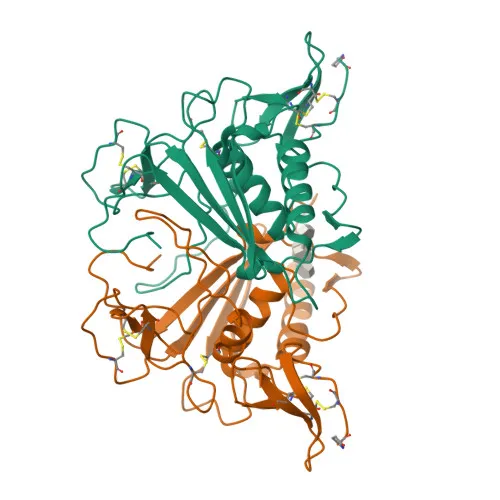
- EpCAM (Epithelial cell adhesion molecule), also known as TACSTD1, TROP1, EGP40, and CD326, is a ~35 kDa type I transmembrane glycoprotein that acts as a homophilic cell adhesion molecule.
- It mediates calcium-independent homophilic interactions between intestinal epithelial cells and intraepithelial lymphocytes, contributing to the immunological barrier at mucosal surfaces and providing a first line of defense against infection.
- EpCAM plays a role in embryonic stem cell (ESC) proliferation and differentiation, and its expression rapidly decreases upon ESC differentiation; it up-regulates the expression of FABP5, MYC, and cyclins A and E.
- The protein localizes to the lateral cell membrane and is enriched at tight junctions, consistent with its role in cell–cell adhesion and epithelial barrier integrity.
- EpCAM is highly and selectively expressed in undifferentiated ESCs and is found on the surface of almost all epithelial cell membranes, but is absent from mesodermal and neural cells; it is also detected on adenocarcinoma cells.
- Post-translational modifications include glycosylation and disulfide bond formation, and EpCAM contains a signal peptide, an extracellular domain with thyroglobulin type-1 repeats, a single transmembrane helix, and a short cytoplasmic tail; it is recognized as a tumor antigen and a disease variant in hereditary nonpolyposis colorectal cancer (HNPCC).
Experimental Guidance and Technical Tips
- The immunogen corresponds to the extracellular domain (amino acids 24–265), which is likely accessible in native conformation; this design may support applications such as WB, IF/ICC, and ELISA.
- For Western blot, the predicted molecular weight is approximately 35 kDa; however, due to glycosylation and other modifications, the observed band may migrate differently. Validate in relevant human, mouse, or rat epithelial cell lysates.
- In IF/ICC, focus on membrane staining with punctate junctional enrichment; appropriate permeabilization is critical. Include appropriate controls (e.g., undifferentiated vs. differentiated ESC lines) to confirm specificity.
- Cross-reactivity with mouse and rat has been reported, but performance in each application should be validated independently in the specific sample system of interest.
CamelBio: Your One-Stop Sourcing Bridge
CamelBio partners with diagnostic manufacturers and research institutes worldwide to provide one-stop access to high-quality IVD raw materials. This EpCAM antibody exemplifies our commitment to supplying validated and optimized immunological reagents for epithelial cell adhesion molecule research. Beyond individual antibodies, we offer validated antibody pairs, bulk ancillary reagents, and custom sourcing for rare targets, helping you accelerate from concept to clinic with confidence.
REQUEST A QUOTE
Our professional team will reply to you within one business day. Please feel free to contact us!