Polyclonal Antibodies
Anti-EIF1AX Polyclonal Antibody for WB, IHC-P, IF/ICC, ELISA - P47813
Item Number : CM0027169
Price varies based on specs and customizations
- Application
- WB, IHC-P, IF/ICC, ELISA
- Cross Reactivity
- Human, Mouse, Rat
- Protein Weight
- 16kDa

Shipping:
Contact us to get shipping details Enjoy On-time Dispatch Guarantee.
Why Choose Us
Easy ordering process, quality products, and dedicated support for your business success.
Core Product Specifications and Parameters
| Parameter | Details |
|---|---|
| Product Name | EIF1AX Rabbit pAb |
| Remarks/Alias | EIF1A, EIF4C, eIF-1A, eIF-4C, EIF1AP1, EIF1AX |
| Species | Human |
| GeneID (Human) | 1964 |
| GeneID | 1964 |
| Immunogen | Recombinant fusion protein containing a sequence corresponding to amino acids 10-144 of human EIF1AX (NP_001403.1) |
| Source | Rabbit |
| Category | Polyclonal Antibodies |
| Application | WB, IHC-P, IF/ICC, ELISA |
| Cross Reactivity | Human, Mouse, Rat |
| SWISS | P47813 |
| Protein Weight | 16kDa |
| Shipping | Ice bag |
Biological Background: EIF1AX Function and Localization
- EIF1AX is the human X-chromosomal gene encoding eukaryotic translation initiation factor 1A, also known as eIF-4C.
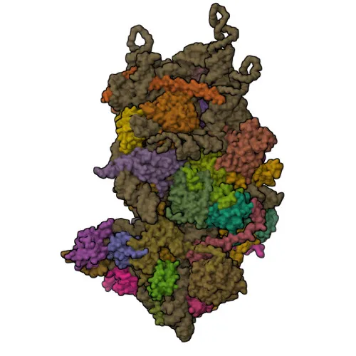
- Functions as a component of the 43S pre-initiation complex (43S PIC) that binds mRNA and scans the 5' UTR to locate the start codon. Together with EIF1, it facilitates mRNA scanning, start codon recognition, and promotes assembly of the 48S initiation complex, while also dissociating aberrant complexes. Related references: PMID:9732867
- After start codon location, cooperates with EIF5B to orient the initiator methionine-tRNA for 60S ribosomal subunit joining, forming the 80S initiation complex. Its globular part resides in the A site of the 40S subunit. Related references: PMID:35732735
- Released from the ribosome after 80S complex formation, following GTP hydrolysis by EIF5B, prior to EIF5B release. Related references: PMID:35732735
- Interaction with EIF5 during scanning helps maintain EIF1 within the open 43S PIC; however, unlike yeast orthologs, human EIF1AX does not directly bind EIF1. Related references: PMID:24319994
- Localizes to the cytoplasm, consistent with its role in translation initiation.
- Keywords include: initiation factor, protein biosynthesis, RNA-binding, tRNA-binding, and host-virus interaction.
Experimental Guidance and Technical Tips
- The immunogen covers amino acids 10-144 of human EIF1AX, a region highly conserved across species; consider testing cross-reactivity with other mammals if needed.
- For Western blot, a 15% SDS-PAGE gel is recommended due to the low molecular weight (~16 kDa). Use appropriate loading controls and verify band specificity with knockdown or knockout samples.
- In IHC-P, antigen retrieval may be required; optimize fixation and retrieval conditions for your tissue type. Include positive and negative controls.
- For IF/ICC, permeabilization is critical for nuclear/cytoplasmic targets. Use 0.1-0.3% Triton X-100 and validate the antibody in your cell system.
- The antibody has been reported to recognize EIF1AX in human, mouse, and rat; however, always validate the antibody in the relevant species and experimental context.
CamelBio: Your One-Stop Sourcing Bridge
CamelBio offers diagnostic manufacturers and research labs streamlined access to high-quality IVD raw materials like the EIF1AX polyclonal antibody for translation initiation studies. Leveraging an extensive supplier network, we provide validated antibody pairs, optimized monoclonal/polyclonal antibodies, bulk ancillary reagents, and sourcing support for rare targets—all tailored to accelerate your assay development from concept to clinic.
Product Datasheet
Anti-EIF1AX Polyclonal Antibody for WB, IHC-P, IF/ICC, ELISA - P47813
REQUEST A QUOTE
Our professional team will reply to you within one business day. Please feel free to contact us!