Monoclonal Antibodies
Anti-EGFR Monoclonal Antibody for WB, IF-P, ELISA - P00533
Item Number : CM0004516
Price varies based on specs and customizations
- Application
- WB, IF-P, ELISA
- Cross Reactivity
- Human, Mouse, Rat
- Protein Weight
- 134kDa

Shipping:
Contact us to get shipping details Enjoy On-time Dispatch Guarantee.
Why Choose Us
Easy ordering process, quality products, and dedicated support for your business success.
Core Product Specifications and Parameters
| Parameter | Value |
|---|---|
| Product Name | [KD Validated] EGFR Rabbit mAb |
| Remarks/Alias | ERBB; ERRP; HER1; mENA; ERBB1; PIG61; NISBD2; EGFR |
| Species | Human |
| GeneID (Human) | 1956 |
| GeneID | 1956 |
| Immunogen | Recombinant fusion protein containing a sequence corresponding to amino acids 335-525 of human EGFR (NP_005219.2) |
| Source | Rabbit |
| Category | Monoclonal Antibodies |
| Application | WB, IF-P, ELISA |
| Cross Reactivity | Human, Mouse, Rat |
| SWISS | P00533 |
| Protein Weight | 134kDa |
| Shipping | Ice bag |
Biological Background: EGFR Function and Localization
-
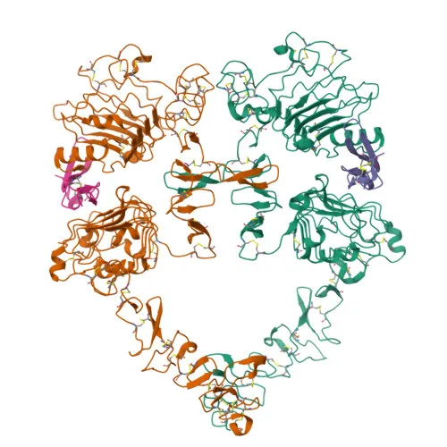
Epidermal growth factor receptor (EGFR) is a receptor tyrosine kinase belonging to the ErbB family, also known as ERBB1/HER1. It binds multiple EGF family ligands including EGF, TGF-α, and HB-EGF, leading to receptor dimerization and autophosphorylation on cytoplasmic tyrosine residues.
Related references: PMID:10805725, PMID:2790960 -
Upon activation, EGFR triggers several major intracellular signaling cascades: RAS-RAF-MEK-ERK, PI3K-AKT, PLCγ-PKC, and STAT modules, thereby regulating cell proliferation, survival, migration, and differentiation. It also activates NF-κB signaling and can directly phosphorylate other targets such as RGS16 and MUC1.
Related references: PMID:27153536, PMID:11116146, PMID:11602604, PMID:11483589 -
Subcellular localization is predominantly at the cell membrane; however, EGFR can also be found in the endoplasmic reticulum, Golgi, endosomes, and nucleus, and a secreted isoform exists.
-
EGFR is a heavily modified glycoprotein with a calculated molecular weight of 134 kDa, featuring key PTMs including phosphorylation, ubiquitination, lipidation, and disulfide bonds.
-
The receptor is ubiquitously expressed in human tissues, and an alternatively spliced isoform 2, which may act as an antagonist, is also detected in ovarian cancers.
-
In addition to its normal physiological roles, EGFR serves as a host cell receptor for hepatitis C virus (HCV) entry, mediating formation of the CD81-CLDN1 complex and enhancing virus-cell membrane fusion.
Experimental Guidance and Technical Tips
- The immunogen corresponds to the extracellular domain (amino acids 335–525) of human EGFR; consider its conservation across species when designing experiments for mouse and rat samples.
- For Western blot, the predicted molecular weight is 134 kDa; use an appropriate gel percentage and optimize transfer conditions for large proteins.
- In immunofluorescence on paraffin sections (IF-P), antigen retrieval may be necessary; validate staining in positive control tissues such as skin or lung.
- For ELISA, test the antibody as a capture or detection reagent after confirming compatibility with your specific assay format.
CamelBio: Your One-Stop Sourcing Bridge
CamelBio connects diagnostic manufacturers, research labs, and clinical facilities with a comprehensive portfolio of IVD raw materials, including this validated anti-EGFR monoclonal antibody for cell signaling and oncology biomarker studies. We offer validated antibody pairs, bulk ancillary reagents, and sourcing of rare targets to support your development pipeline from concept to clinic.
REQUEST A QUOTE
Our professional team will reply to you within one business day. Please feel free to contact us!