Monoclonal Antibodies
Anti-EDG1/HEXIM1 Monoclonal Antibody for WB and ELISA - P21453
Item Number : CM0008606
Price varies based on specs and customizations
- Application
- WB, ELISA
- Cross Reactivity
- Human, Mouse, Rat
- Protein Weight
- 43kDa

Shipping:
Contact us to get shipping details Enjoy On-time Dispatch Guarantee.
Why Choose Us
Easy ordering process, quality products, and dedicated support for your business success.
Core Product Specifications and Parameters
| Parameter | Value |
|---|---|
| Product Name | EDG1/HEXIM1 Rabbit mAb |
| Remarks/Alias | EDG1; S1P1; CD363; ECGF1; EDG-1; CHEDG1; D1S3362; EDG1/HEXIM1 |
| Species | Human |
| Gene ID (Human) | 1901 |
| Gene ID | 1901 |
| Immunogen | Synthetic Peptide| A synthetic peptide corresponding to a sequence within amino acids 1-100 of human EDG1/HEXIM1 (P21453). |
| Source | Rabbit |
| Category | Monoclonal Antibodies |
| Application | WB, ELISA |
| Cross Reactivity | Human, Mouse, Rat |
| SWISS | P21453 |
| Protein Weight | 43kDa |
| Shipping | Ice bag |
Biological Background: EDG1/HEXIM1 Function and Localization
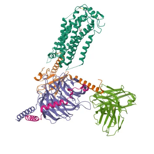
- EDG1/HEXIM1, also known as sphingosine 1-phosphate receptor 1 (S1PR1), endothelial differentiation G protein-coupled receptor 1 (EDG1), CD363, and S1P1, is a member of the G protein-coupled receptor family.
- It serves as a receptor for the bioactive lipid sphingosine 1-phosphate (S1P), coupling to G(i) proteins to activate downstream signaling pathways involving RAC1, SRC, PTK2/FAK1, and MAP kinases, thereby regulating cell migration and actin cytoskeleton reorganization.
- The receptor plays critical roles in angiogenesis and vascular maturation: it promotes endothelial cell migration and inhibits excessive sprouting angiogenesis during blood vessel development.
- EDG1/HEXIM1 is essential for normal embryonic heart development and cardiac morphogenesis, and it is required for the egress of mature T-cells from the thymus into peripheral lymphoid organs.
- Subcellularly, it localizes to the cell membrane, endosomes, and membrane rafts, and undergoes post-translational modifications including acetylation, palmitoylation, and phosphorylation.
- The protein is predominantly expressed in endothelial cells, with lower levels in vascular smooth muscle cells, fibroblasts, melanocytes, and epithelioid-derived cells, and it has a molecular weight of approximately 43 kDa.
Experimental Guidance and Technical Tips
- The immunogen corresponds to the N-terminal extracellular domain (amino acids 1–100), which may contribute to the antibody's specificity for the native receptor conformation; consider using this antibody to detect the receptor in denaturing conditions such as Western blotting.
- Validation of the antibody in ELISA suggests it can recognize the linear epitope; consider optimizing the coating antigen concentration and detection conditions for your specific assay format.
- The antibody is reported to cross-react with mouse and rat EDG1/HEXIM1; perform titration experiments to determine the optimal working dilution for these species and applications.
- Since the receptor is a multi-pass membrane protein with a molecular weight of ~43 kDa, consider including a reducing agent and boiling step in your sample preparation for Western blotting, and confirm band size with appropriate molecular weight markers.
CamelBio: Your One-Stop Sourcing Bridge
CamelBio offers a streamlined sourcing solution for diagnostic manufacturers and research institutions, providing reliable access to specialty reagents like this anti-EDG1/HEXIM1 monoclonal antibody. Our comprehensive portfolio includes validated antibody pairs, optimized monoclonal and polyclonal antibodies, and bulk ancillary reagents, all supported by expert technical guidance to accelerate your assay development. For researchers investigating GPCR signaling, angiogenesis, or immune cell trafficking, CamelBio serves as a trusted partner for procuring high-quality raw materials efficiently.
REQUEST A QUOTE
Our professional team will reply to you within one business day. Please feel free to contact us!