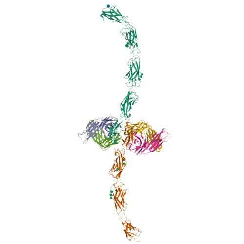
Monoclonal Antibodies
Anti-E-Cadherin Rabbit Monoclonal Antibody for WB, IF/ICC, IF-P, IHC-P, ELISA - P12830
Item Number : CM0002230
Price varies based on specs and customizations
- Application
- WB, IF/ICC, IF-P, IHC-P, ELISA
- Cross Reactivity
- Human, Mouse, Rat
- Protein Weight
- 97kDa

Shipping:
Contact us to get shipping details Enjoy On-time Dispatch Guarantee.
Why Choose Us
Easy ordering process, quality products, and dedicated support for your business success.
Core Product Specifications and Parameters
| Parameter | Value |
|---|---|
| Product Name | E-Cadherin Rabbit mAb |
| Remarks/Alias | UVO; CDHE; ECAD; LCAM; Arc-1; BCDS1; CD324; E-Cadherin |
| Species | Human |
| GeneID | 999 |
| Immunogen | Synthetic peptide corresponding to a sequence within amino acids 701-800 of human E-Cadherin (NP_004351.1) |
| Source | Rabbit |
| Category | Monoclonal Antibodies |
| Application | WB, IF/ICC, IF-P, IHC-P, ELISA |
| Cross Reactivity | Human, Mouse, Rat |
| SWISS | P12830 |
| Protein Weight | 97kDa |
| Shipping | Ice bag |
Biological Background: E-Cadherin Function and Localization
-
Cadherin-1 (E-cadherin) is a calcium-dependent cell adhesion protein that mediates homophilic interactions to connect cells, contributing to the sorting of heterogeneous cell types (PubMed:11976333).
Related references: PMID:11976333 -
E-cadherin is involved in regulating cell-cell adhesions, mobility, and proliferation of epithelial cells (PubMed:11976333).
Related references: PMID:11976333 -
It plays a role in the early stages of desmosome formation by facilitating the recruitment of DSG2 and DSP to desmosome plaques (PubMed:29999492).
Related references: PMID:29999492 -
Functions as a potent invasive suppressor and serves as a ligand for integrin alpha-E/beta-7.
-
The cytoplasmic tail fragment E-Cad/CTF2 promotes non-amyloidogenic degradation of Abeta precursors, inhibiting APP C99 and C83 production.
-
Acts as a receptor for Listeria monocytogenes internalin A, promoting bacterial uptake.
-
Subcellularly localized to adherens junctions, desmosomes, cell membrane, endosome, Golgi, and can be secreted.
-
Expressed in epithelial tissues including skin, esophageal epithelium, and liver; also found in granuloma macrophages.
Experimental Guidance and Technical Tips
- For Western blot (WB), under denaturing conditions, a band around 97 kDa is expected. Include appropriate loading controls and validate in your sample system.
- For immunohistochemistry (IHC-P), optimize antigen retrieval as E-cadherin is a transmembrane protein. Test on skin or esophageal epithelium as positive control tissues.
- The immunogen is a synthetic peptide from the cytoplasmic domain (aa 701-800); it may detect both full-length and cleaved fragments. Consider this when analyzing bands of different sizes.
- For immunofluorescence (IF), perform fixation and permeabilization steps carefully. Co-staining with junctional markers may confirm proper localization.
- Cross-reactivity with mouse and rat is indicated; perform appropriate species-specific controls when using these models.
CamelBio: Your One-Stop Sourcing Bridge
CamelBio is your one-stop IVD raw-material sourcing partner, dedicated to supporting diagnostic manufacturers, laboratories, and research institutes from concept to clinic. This anti-E-Cadherin monoclonal antibody is ideal for studies in cell adhesion, epithelial-mesenchymal transition, and cancer biology. We offer a comprehensive portfolio including validated antibody pairs, bulk ancillary reagents, and customized sourcing for rare targets, ensuring all your E-Cadherin research and development needs are met with efficiency and quality.
Product Datasheet
Anti-E-Cadherin Rabbit Monoclonal Antibody for WB, IF/ICC, IF-P, IHC-P, ELISA - P12830
REQUEST A QUOTE
Our professional team will reply to you within one business day. Please feel free to contact us!