Polyclonal Antibodies
Anti-E-Cadherin Polyclonal Antibody for WB, IHC-P, IF/ICC, ELISA - P12830
Item Number : CM0014358
Price varies based on specs and customizations
- Applications
- WB, IHC-P, IF/ICC, ELISA
- Cross Reactivity
- Human, Mouse, Rat
- Predicted Molecular Weight
- 97kDa

Shipping:
Contact us to get shipping details Enjoy On-time Dispatch Guarantee.
Why Choose Us
Easy ordering process, quality products, and dedicated support for your business success.
Core Product Specifications and Parameters
| Parameter | Value |
|---|---|
| Product Name | E-Cadherin Rabbit pAb |
| Remarks/Alias | UVO; CDHE; ECAD; LCAM; Arc-1; BCDS1; CD324; E-Cadherin |
| Species | Human |
| GeneID (human) | 999 |
| GeneID | 999 |
| Immunogen | A synthetic peptide corresponding to a sequence within amino acids 373-622 of human E-Cadherin (NP_004351.1). |
| Source | Rabbit |
| Category | Polyclonal Antibodies |
| Application | WB, IHC-P, IF/ICC, ELISA |
| Cross Reactivity | Human, Mouse, Rat |
| SWISS | P12830 |
| Protein Weight | 97kDa |
| Shipping | Ice bag |
Biological Background: E-Cadherin Function and Localization
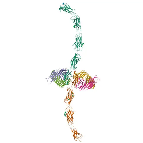
- E-Cadherin (Cadherin-1, also known as CAM 120/80, Epithelial cadherin, Uvomorulin) is a classical cadherin encoded by the CDH1 gene. It belongs to the cadherin superfamily of calcium-dependent cell adhesion molecules.
- It mediates homophilic cell-cell adhesion at adherens junctions, which is essential for maintaining epithelial tissue integrity and architecture. Related references: PMID:11976333
- E-Cadherin regulates cell sorting, mobility, and proliferation. Through interaction with AMOTL2, it anchors radial actin fibers to junctional complexes and transduces contractile forces.
- It facilitates early desmosome assembly by recruiting desmoglein-2 (DSG2) and desmoplakin (DSP) to desmosomal plaques. Related references: PMID:29999492
- The protein localizes to the cell membrane, adherens junctions, and desmosomes; it also cycles through the endosomal–Golgi network and can be secreted.
- Post-translational modifications include cleavage, phosphorylation, ubiquitination, disulfide bond formation, and N‑glycosylation.
- Expressed in epithelial tissues (skin, liver, esophageal epithelium) and in granuloma macrophages. Related references: PMID:27760340 PMID:22294297 PMID:24220297 PMID:3263290
- Serves as a receptor for Listeria monocytogenes internalin A, promoting bacterial entry into host cells.
Experimental Guidance and Technical Tips
- The immunogen covers amino acids 373–622, which lies within the extracellular cadherin repeat region; consider that the antibody may recognize both full-length E-Cadherin and proteolytic fragments.
- Predicted molecular weight is ~97 kDa; however, due to extensive glycosylation and propeptide processing, the mature protein typically migrates at 120–135 kDa in SDS-PAGE. Validate in your sample system.
- For IHC-P, perform heat-induced antigen retrieval in citrate buffer (pH 6.0) and optimize primary antibody dilution.
- Cross-reactivity is expected with mouse and rat based on sequence homology; verify for your specific application.
- Titrate the antibody for ELISA and IF/ICC to determine optimal working concentrations.
CamelBio: Your One-Stop Sourcing Bridge
CamelBio offers comprehensive access to IVD raw materials, supporting diagnostic manufacturers and research laboratories from concept to clinical application. For E-Cadherin-targeted assays, we can supply validated antibody pairs, bulk monoclonal/polyclonal reagents, and ancillary assay components. Our sourcing platform also handles custom requirements for rare or challenging targets, providing a seamless procurement experience.
Product Datasheet
Anti-E-Cadherin Polyclonal Antibody for WB, IHC-P, IF/ICC, ELISA - P12830
REQUEST A QUOTE
Our professional team will reply to you within one business day. Please feel free to contact us!