Polyclonal Antibodies
Anti-DUSP9 (MKP-4) Polyclonal Antibody for WB, IF/ICC, ELISA - Q99956
Item Number : CM0025603
Price varies based on specs and customizations
- Application
- WB, IF/ICC, ELISA
- Cross Reactivity
- Human, Mouse
- Protein Weight
- 42kDa

Shipping:
Contact us to get shipping details Enjoy On-time Dispatch Guarantee.
Why Choose Us
Easy ordering process, quality products, and dedicated support for your business success.
Core Product Specifications and Parameters
| Parameter | Value |
|---|---|
| Product Name | DUSP9 Rabbit pAb |
| Remarks/Alias | MKP4, MKP-4, DUSP9 |
| Species | Human |
| Gene ID | 1852 |
| Immunogen | Recombinant fusion protein containing a sequence corresponding to amino acids 1-200 of human DUSP9 (NP_001386.1) |
| Source | Rabbit |
| Category | Polyclonal Antibodies |
| Application | WB, IF/ICC, ELISA |
| Cross Reactivity | Human, Mouse |
| SWISS | Q99956 |
| Protein Weight | 42kDa |
| Shipping | Ice bag |
Biological Background: DUSP9 Function and Localization
- DUSP9 encodes a dual specificity protein phosphatase that belongs to the MAP kinase phosphatase (MKP) family, specifically designated as MKP-4.
- It dephosphorylates and inactivates MAP kinases, with a strong preference for the ERK (extracellular signal-regulated kinase) subfamily, thereby negatively regulating ERK signaling cascades.
- DUSP9 is localized in the cytoplasm, where it encounters activated ERK kinases to mediate signal attenuation.
- The protein is classified as a phosphoprotein, indicating that its activity or stability may be regulated by phosphorylation.
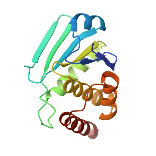
- Structural studies (keyword: 3D-structure) have determined the three-dimensional arrangement of DUSP9, providing insights into its catalytic mechanism and substrate recognition.
- Keywords such as Hydrolase, Protein phosphatase, and Reference proteome highlight its enzymatic function and curated annotation status.
Experimental Guidance and Technical Tips
- The immunogen corresponds to the N-terminal region (aa 1–200) of human DUSP9; the resulting polyclonal antibody may recognize both phosphorylated and unphosphorylated forms. Consider this when probing samples with varying phosphatase activity.
- For western blot (WB), a band at ~42 kDa is expected in human and mouse lysates. Verify specificity using appropriate controls (e.g., ERK activation or knockdown models).
- In immunofluorescence (IF/ICC), anticipate cytoplasmic staining consistent with DUSP9 localization. Co-staining with ERK markers can validate functional context.
- ELISA applications require optimization of coating antigen and detection conditions; cross-reactivity with mouse DUSP9 enables comparative studies.
- When working with phosphatase inhibitors, be aware that they may alter DUSP9 phosphorylation status and antibody binding.
CamelBio: Your One-Stop Sourcing Bridge
As a dedicated IVD raw-material sourcing partner, CamelBio supports diagnostic manufacturers and researchers with validated antibody pairs and optimized polyclonal antibodies like this anti-DUSP9 pAb. We also supply bulk ancillary reagents and excel in sourcing rare targets, ensuring you have the tools to advance ERK signaling studies and phosphatase-based diagnostics from concept to clinic.
Product Datasheet
Anti-DUSP9 (MKP-4) Polyclonal Antibody for WB, IF/ICC, ELISA - Q99956
REQUEST A QUOTE
Our professional team will reply to you within one business day. Please feel free to contact us!