Polyclonal Antibodies
Anti-DRD1 Rabbit Polyclonal Antibody for WB and ELISA - P21728
Item Number : CM0028119
Price varies based on specs and customizations
- Application
- WB, ELISA
- Cross Reactivity
- Rat
- Protein Weight
- 49kDa

Shipping:
Contact us to get shipping details Enjoy On-time Dispatch Guarantee.
Why Choose Us
Easy ordering process, quality products, and dedicated support for your business success.
Core Product Specifications and Parameters
| Parameter | Value |
|---|---|
| Product Name | DRD1 Rabbit pAb |
| Remarks/Alias | DADR; DRD1A; DRD1 |
| Species | Human |
| GeneID (Human) | 1812 |
| GeneID | 1812 |
| Immunogen | Recombinant fusion protein containing a sequence corresponding to amino acids 338-446 of human DRD1 (NP_000785.1) |
| Source | Rabbit |
| Category | Polyclonal Antibodies |
| Application | WB, ELISA |
| Cross Reactivity | Rat |
| SWISS | P21728 |
| Protein Weight | 49kDa |
| Shipping | Ice bag |
Biological Background: DRD1 Function and Localization
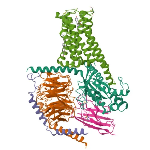
- DRD1 (D(1A) dopamine receptor) is a G protein-coupled receptor that mediates dopamine signaling by activating adenylyl cyclase through G proteins, increasing intracellular cAMP levels.
- The receptor forms heterotetramers with DRD3, which enhances beta-arrestin recruitment and contributes to the regulation of locomotor activity.
- DRD1 is primarily expressed in the central nervous system, with high levels detected in the caudate, nucleus accumbens, and olfactory tubercle.
- Subcellularly, DRD1 localizes to the cell membrane, endoplasmic reticulum membrane, and specific neuronal compartments including the cilium membrane, dendrites, and dendritic spines.
- As a transmembrane protein, it undergoes post-translational modifications such as glycosylation, palmitoylation, and formation of disulfide bonds, which are critical for proper folding and membrane trafficking.
- The receptor belongs to the dopamine receptor family and is a model system for understanding GPCR signaling and synaptic transmission.
Experimental Guidance and Technical Tips
- The immunogen corresponds to the C-terminal region (amino acids 338-446) of human DRD1, suggesting the antibody will recognize epitopes in this domain; consider this for experimental design.
- For Western blot, a band around 49 kDa is expected; use a 10% SDS-PAGE gel and validate in brain tissue lysates from human or rat origin, given the reported cross-reactivity.
- In ELISA applications, pair with a suitable detection antibody and include appropriate controls using recombinant DRD1 protein or peptide standards.
- As a polyclonal antibody, it may show additional bands in WB; optimize blocking and dilution conditions to minimize non-specific binding.
CamelBio: Your One-Stop Sourcing Bridge
CamelBio serves as a comprehensive sourcing partner for IVD raw materials, connecting diagnostic manufacturers and research labs with validated reagents. For neuroscience and GPCR signaling research, we offer this anti-DRD1 polyclonal antibody and can supply matched antibody pairs, recombinant proteins, and ancillary reagents to streamline your assay development. Leverage our one-stop platform to source rare target reagents and accelerate your diagnostic projects from concept to clinic.
REQUEST A QUOTE
Our professional team will reply to you within one business day. Please feel free to contact us!