Polyclonal Antibodies
Anti-DP1/TFDP1 Rabbit Polyclonal Antibody for IHC-P and ELISA - Q14186
Item Number : CM0013111
Price varies based on specs and customizations
- Application
- IHC-P, ELISA
- Cross Reactivity
- Mouse, Rat
- Protein Weight
- 45kDa

Shipping:
Contact us to get shipping details Enjoy On-time Dispatch Guarantee.
Why Choose Us
Easy ordering process, quality products, and dedicated support for your business success.
Core Product Specifications and Parameters
| Parameter | Value |
|---|---|
| Product Name | DP1/TFDP1 Rabbit pAb |
| Remarks/Alias | DP1; DILC; Dp-1; DRTF1; DP1/TFDP1 |
| Species | Human |
| GeneID (Human) | 7027 |
| Immunogen | Recombinant fusion protein containing a sequence corresponding to amino acids 1-290 of human DP1/TFDP1 (NP_009042.1) |
| Source | Rabbit |
| Category | Polyclonal Antibodies |
| Application | IHC-P, ELISA |
| Cross Reactivity | Mouse, Rat |
| SWISS | Q14186 |
| Protein Weight | 45kDa |
| Shipping | Ice bag |
Biological Background: DP1/TFDP1 Function and Localization
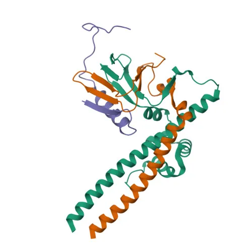
- DP1/TFDP1, also known as DRTF1-polypeptide 1 or E2F dimerization partner 1, is a transcription factor that dimerizes with E2F family members to regulate gene expression.
- It stimulates E2F-dependent transcription by binding cooperatively to the E2 recognition site 5'-TTTC[CG]CGC-3' in promoters of genes involved in cell cycle regulation and DNA replication. Related references: PMID:7739537 PMID:8405995
- The E2F1:DP complex mediates both cell proliferation and apoptosis, highlighting a dual role in cell fate decisions.
- DP1/TFDP1 blocks adipocyte differentiation by repressing CEBPA binding to its target gene promoters. Related references: PMID:20176812
- Subcellularly, DP1/TFDP1 localizes to both the nucleus and cytoplasm, consistent with its transcriptional regulatory functions.
- Tissue expression analysis reveals highest levels in muscle, with additional expression in brain, placenta, liver, and kidney; lower levels in lung and pancreas; and absence in heart.
- Post-translational modifications include acetylation and phosphorylation, which may modulate its activity and interactions, and it is subject to ubiquitin conjugation.
Experimental Guidance and Technical Tips
- The immunogen comprising amino acids 1-290 suggests the antibody recognizes an N-terminal epitope, suitable for detecting both native and denatured DP1/TFDP1; consider testing in western blotting under reducing and non-reducing conditions if needed.
- For immunohistochemistry (IHC-P), optimize antigen retrieval protocols (e.g., heat-induced epitope retrieval in citrate buffer) and validate staining specificity using appropriate positive and negative tissue controls.
- In ELISA applications, consider using recombinant DP1/TFDP1 protein as a standard to assess antibody sensitivity and linear range.
- Given cross-reactivity with mouse and rat, validate the antibody in the relevant species and tissue types to confirm reactivity and specificity prior to experimental use.
CamelBio: Your One-Stop Sourcing Bridge
CamelBio offers a comprehensive portfolio of IVD raw materials, including validated antibody pairs, optimized monoclonal and polyclonal antibodies, and bulk ancillary reagents, supporting diagnostic manufacturers and researchers from concept to clinic. This anti-DP1/TFDP1 antibody aids in studying E2F-mediated transcription, cell cycle control, and cancer biology, and CamelBio can assist with sourcing related rare targets, custom antibody development, and procurement of matched reagents for your assay pipeline. Our one-stop supply model ensures reliable access to critical components, enabling you to accelerate your diagnostic development with confidence.
Product Datasheet
Anti-DP1/TFDP1 Rabbit Polyclonal Antibody for IHC-P and ELISA - Q14186
REQUEST A QUOTE
Our professional team will reply to you within one business day. Please feel free to contact us!