Polyclonal Antibodies
Anti-DP1/TFDP1 Polyclonal Antibody for WB, IHC-P, ELISA - Q14186
Item Number : CM0026491
Price varies based on specs and customizations
- Application
- WB, IHC-P, ELISA
- Cross Reactivity
- Human, Mouse
- Protein Weight
- 45kDa

Shipping:
Contact us to get shipping details Enjoy On-time Dispatch Guarantee.
Why Choose Us
Easy ordering process, quality products, and dedicated support for your business success.
Core Product Specifications and Parameters
| Parameter | Value |
|---|---|
| Product Name | DP1/TFDP1 Rabbit pAb |
| Remarks/Alias | DP1; DILC; Dp-1; DRTF1; DP1/TFDP1 |
| Species | Human |
| GeneID (Human) | 7027 |
| GeneID | 7027 |
| Immunogen | Recombinant fusion protein containing a sequence corresponding to amino acids 1-290 of human DP1/TFDP1 (NP_009042.1). |
| Source | Rabbit |
| Category | Polyclonal Antibodies |
| Application | WB, IHC-P, ELISA |
| Cross Reactivity | Human, Mouse |
| SWISS | Q14186 |
| Protein Weight | 45kDa |
| Shipping | Ice bag |
Biological Background: DP1/TFDP1 Function and Localization
-
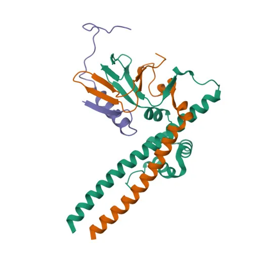
Transcription factor Dp-1 (TFDP1) is a key component of the E2F transcription factor complex, serving as the obligate dimerization partner for E2F family proteins.
-
DP1 binds DNA cooperatively with E2F through the E2 recognition site 5'-TTTC[CG]CGC-3' found in promoters of genes involved in cell cycle progression and DNA replication.
Related references: PMID:7739537, PMID:8405995
-
The E2F1:DP complex appears to mediate both cell proliferation and apoptosis, highlighting its dual role in cell fate decisions.
-
DP1 blocks adipocyte differentiation by repressing CEBPA binding to its target gene promoters.
Related references: PMID:20176812
-
DP1 localizes to both the nucleus and cytoplasm, enabling its transcriptional regulatory functions and potential cytoplasmic roles.
-
Tissue expression is highest in muscle, with moderate levels in brain, placenta, liver, and kidney; lower in lung and pancreas; and undetectable in heart.
-
DP1 undergoes post-translational modifications including acetylation, phosphorylation, and ubiquitination, which modulate its stability and activity.
Experimental Guidance and Technical Tips
- The immunogen corresponds to the N-terminal region (aa 1-290), so the antibody is expected to recognize full-length DP1 (~45 kDa). Validate detection in relevant cell or tissue lysates.
- For Western blotting, optimize antibody dilution; a band around 45 kDa is anticipated. Use appropriate loading controls.
- For immunohistochemistry (paraffin), standard antigen retrieval methods are recommended; titrate antibody for optimal signal-to-noise ratio.
- This antibody cross-reacts with mouse, making it suitable for comparative studies. For other species, perform sequence alignment and test experimentally.
- In ELISA applications, consider using a validated antigen as positive control.
CamelBio: Your One-Stop Sourcing Bridge
CamelBio supports diagnostic manufacturers and research institutions with comprehensive access to IVD raw materials, technical services, and consulting—from concept to clinic. For the DP1/TFDP1 target and cell cycle research, we supply optimized monoclonal and polyclonal antibodies, validated antibody pairs, bulk ancillary reagents, and rare target sourcing, ensuring a seamless procurement experience for your assay development needs.
REQUEST A QUOTE
Our professional team will reply to you within one business day. Please feel free to contact us!