Polyclonal Antibodies
Anti-DNAJA3 Polyclonal Antibody for WB, ELISA - Q96EY1
Item Number : CM0029837
Price varies based on specs and customizations
- Application
- WB, ELISA
- Cross Reactivity
- Human, Mouse, Rat
- Protein Weight
- 52kDa

Shipping:
Contact us to get shipping details Enjoy On-time Dispatch Guarantee.
Why Choose Us
Easy ordering process, quality products, and dedicated support for your business success.
Core Product Specifications and Parameters
| Parameter | Value |
|---|---|
| Product Name | DNAJA3 Rabbit pAb |
| Remarks/Alias | TID1; HCA57; hTID-1; DNAJA3 |
| Species | Human |
| Gene ID (Human) | 9093 |
| Gene ID | 9093 |
| Immunogen | Recombinant fusion protein containing a sequence corresponding to amino acids 221-480 of human DNAJA3 (NP_005138.3) |
| Source | Rabbit |
| Category | Polyclonal Antibodies |
| Application | WB, ELISA |
| Cross Reactivity | Human, Mouse, Rat |
| SWISS | Q96EY1 |
| Protein Weight | 52kDa |
| Shipping | Ice bag |
Biological Background: DNAJA3 Function and Localization
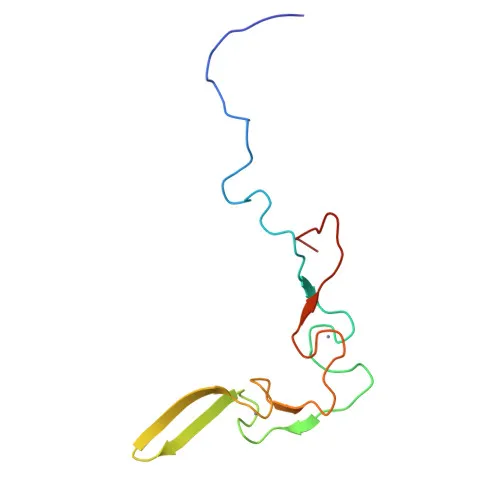
- DNAJA3, also known as TID1, hTid-1, and HCA57, is a mitochondrial member of the DnaJ/Hsp40 chaperone family, characterized by a zinc-finger domain and metal-binding properties.
- Encoded by the DNAJA3 gene, two main alternatively spliced isoforms exist: isoform 1 and isoform 2, which play opposing roles in apoptosis regulation.
- Isoform 1 increases apoptosis triggered by TNF and DNA-damaging agents like mitomycin C, while isoform 2 suppresses apoptosis, demonstrating functional duality.
- The protein modulates apoptotic signal transduction by affecting cytochrome c release from mitochondria and caspase-3 activation, without affecting caspase-8.
- Isoform 2 may serve as an effector of the MUSK signaling pathway, contributing to neuromuscular junction development.
- Subcellular localization includes mitochondrial matrix, cytoplasm, cytosol, and postsynaptic cell membrane, highlighting its dynamic intracellular distribution.
- Widely expressed in human tissues, with highest levels detected in heart, liver, lung, and skeletal muscle; expression has also been observed in keratinocytes, with altered distribution in basal cell carcinomas. Related references: PMID:9683573 PMID:12783860
- Keywords such as acetylation, phosphorylation, methylation, and proteomics identification suggest multiple post-translational modifications that may regulate its function and stability.
Experimental Guidance and Technical Tips
- The immunogen used for this antibody spans amino acids 221-480 of human DNAJA3, covering a significant portion of the protein and expected to provide good specificity. Validate in the relevant sample system to confirm reactivity.
- For Western blotting, a band at approximately 52 kDa is expected. Consider using mitochondrial-enriched lysates or cell lines with known DNAJA3 expression for optimal detection.
- The antibody has been validated for ELISA; appropriate serum or cell culture supernatant dilutions should be experimentally determined.
- Given the cross-reactivity with mouse and rat, this antibody can be employed in comparative studies across these species; however, sequence homology should be verified for each model.
CamelBio: Your One-Stop Sourcing Bridge
CamelBio is your integrated one-stop partner for IVD raw-material procurement, providing validated antibodies, optimized monoclonal/polyclonal reagents, bulk ancillary supplies, and expert sourcing for rare targets. This high-quality rabbit polyclonal antibody against DNAJA3 supports apoptosis, mitochondrial biology, and neuromuscular junction research, enabling your diagnostic assay development from concept to clinic. Explore our comprehensive portfolio to accelerate your project timelines.
REQUEST A QUOTE
Our professional team will reply to you within one business day. Please feel free to contact us!