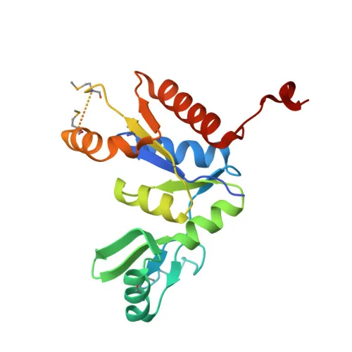
Polyclonal Antibodies
Anti-DLG1 Polyclonal Antibody for WB, IF/ICC, ELISA - Q12959
Item Number : CM0026706
Price varies based on specs and customizations
- Application
- WB, IF/ICC, ELISA
- Cross Reactivity
- Human, Mouse, Rat
- Protein Weight
- 100kDa

Shipping:
Contact us to get shipping details Enjoy On-time Dispatch Guarantee.
Why Choose Us
Easy ordering process, quality products, and dedicated support for your business success.
Core Product Specifications and Parameters
| Parameter | Specification |
|---|---|
| Product Name | DLG1 Rabbit pAb |
| Remarks/Alias | hdlg; DLGH1; SAP97; SAP-97; DLG1 |
| Species | Human |
| Gene ID (Human) | 1739 |
| Gene ID | 1739 |
| Immunogen | Recombinant fusion protein containing a sequence corresponding to amino acids 1-190 of human DLG1 (NP_004078.2). |
| Source | Rabbit |
| Category | Polyclonal Antibodies |
| Application | WB, IF/ICC, ELISA |
| Cross Reactivity | Human, Mouse, Rat |
| SWISS | Q12959 |
| Protein Weight | 100kDa |
| Shipping | Ice bag |
Biological Background: DLG1 Function and Localization
- DLG1, also known as synapse-associated protein 97 (SAP97) or hDlg, is a membrane-associated guanylate kinase (MAGUK) scaffolding protein that organizes multiprotein complexes at cellular junctions.
- It plays essential roles in cell polarity by recruiting channels, receptors, and signaling molecules to discrete plasma membrane domains in polarized cells, and promotes epithelial barrier function through cell-cell adhesion maintenance (By similarity).
- DLG1 regulates cardiac myocyte excitability by modulating the functional expression of Kv4 channels and also acts as a functional regulator of the Kv1.5 potassium channel.
- In hippocampal neurons, it participates in long-term depression by recruiting ADAM10 to the plasma membrane, a process critical for synaptic plasticity. Related references: PMID:23676497
- Subcellular localization includes the basolateral and apical cell membrane, endoplasmic reticulum, postsynaptic density, synapse, and cell-cell junctions, reflecting its scaffolding roles.
- Tissue specificity: abundantly expressed in atrial myocardium (protein level) and detected in lung fibroblasts, cervical epithelia, B-cells, and brain. Related references: PMID:23676497
- Keywords such as alternative splicing, phosphoprotein, and host-virus interaction highlight its regulatory complexity and involvement in diverse cellular processes.
Experimental Guidance and Technical Tips
- The immunogen (amino acids 1–190) corresponds to the N-terminal region of human DLG1. Because alternative splicing generates multiple isoforms, consider sequence alignment of this region with your target isoform(s) before use.
- For Western blot, a band near 100 kDa is expected. Due to potential isoform variation or post-translational modifications, additional bands may be observed; optimize antibody dilution and validate in your sample system.
- When using immunofluorescence (IF/ICC), check the subcellular distribution (e.g., membrane, junctions, or synapse) consistent with the known localization; include appropriate controls for specificity.
- Cross-reactivity with mouse and rat is indicated; however, always verify performance in your species and tissue of interest by including positive and negative controls.
CamelBio: Your One-Stop Sourcing Bridge
CamelBio connects diagnostic manufacturers, CROs, and research laboratories with a broad portfolio of IVD raw materials. This anti-DLG1 polyclonal antibody supports investigations in cell polarity, neuroscience, and cardiac physiology. Beyond single antibody orders, CamelBio offers validated antibody pairs, bulk ancillary reagents, and custom sourcing for rare-target raw materials, streamlining your development from concept to clinic.
REQUEST A QUOTE
Our professional team will reply to you within one business day. Please feel free to contact us!