Monoclonal Antibodies
Anti-DLAT Rabbit Monoclonal Antibody for WB, IF/ICC, IHC-P, ELISA - P10515
Item Number : CM0010793
Price varies based on specs and customizations
- Application
- WB, IF/ICC, IHC-P, ELISA
- Cross Reactivity
- Human, Mouse, Rat
- Protein Weight
- 69 kDa

Shipping:
Contact us to get shipping details Enjoy On-time Dispatch Guarantee.
Why Choose Us
Easy ordering process, quality products, and dedicated support for your business success.
Core Product Specifications and Parameters
| Parameter | Value |
|---|---|
| Product Name | DLAT Rabbit mAb |
| Remarks/Alias | E2, PBC, DLTA, PDCE2, PDC-E2 |
| Species | Human |
| GeneID (Human) | 1737 |
| GeneID | 1737 |
| Immunogen | Recombinant fusion protein containing a sequence corresponding to amino acids 87-270 of human DLAT (NP_001922.2) |
| Source | Rabbit |
| Category | Monoclonal Antibodies |
| Application | WB, IF/ICC, IHC-P, ELISA |
| Cross Reactivity | Human, Mouse, Rat |
| SWISS | P10515 |
| Protein Weight | 69 kDa |
| Shipping | Ice bag |
Biological Background: DLAT Function and Localization
- DLAT encodes the dihydrolipoamide acetyltransferase component (E2) of the mitochondrial pyruvate dehydrogenase (PDH) complex, which catalyzes pyruvate to acetyl-CoA conversion, linking glycolysis and the TCA cycle (Probable).
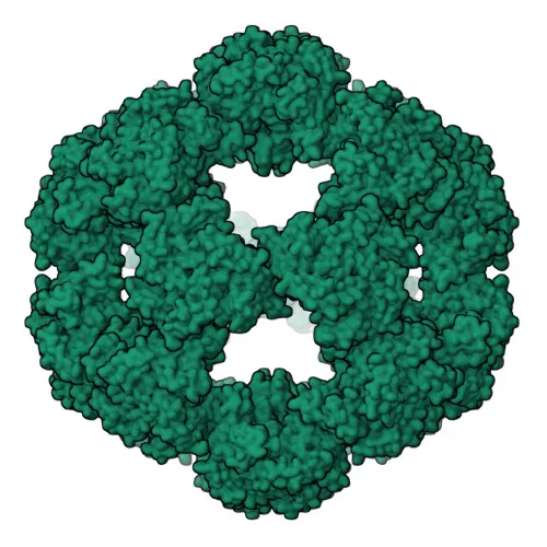
- The PDH complex contains multiple copies of three enzymes: E1 (pyruvate dehydrogenase), E2 (dihydrolipoamide acetyltransferase), and E3 (dihydrolipoamide dehydrogenase). DLAT constitutes the E2 core, accepting acetyl groups from the lipoyl moiety of E1 and transferring them to coenzyme A to form acetyl-CoA (Probable).
- Known aliases for DLAT include PDC-E2, PDCE2, DLTA, E2, and PBC, the latter reflecting its identification as the major autoantigen in primary biliary cirrhosis (M2 antigen complex 70 kDa subunit).
- Subcellularly, DLAT localizes to the mitochondrial matrix, as indicated by its SUBCELLULAR LOCATION annotation and the presence of a mitochondrial transit peptide.
- Post-translational modifications such as acetylation, lipoylation, and phosphorylation are noted in UniProt keywords, highlighting the complex regulation of the PDH complex.
- Proteomics identification and reference proteome status confirm DLAT's robust detection in proteomic studies, consistent with its role as a key metabolic enzyme.
Experimental Guidance and Technical Tips
- The immunogen covers amino acids 87-270, which includes the lipoyl-binding domain; therefore, this antibody is expected to detect both native and recombinant DLAT. For WB, the expected band size is approximately 69 kDa; using mitochondrial-enriched samples may improve signal specificity.
- In IF/ICC, anticipate mitochondrial staining; co-localization with organelle markers (e.g., MitoTracker) is recommended for validation.
- The antibody is cross-reactive with mouse and rat; verify sequence homology in the immunogen region if species-specific detection is critical.
- For IHC-P, consider antigen retrieval and dilution optimization. Use positive control tissues expressing high levels of DLAT (e.g., liver, heart) to confirm reactivity.
- For ELISA, ensure the recombinant protein or peptide antigen matches the immunogen sequence to avoid epitope masking.
CamelBio: Your One-Stop Sourcing Bridge
CamelBio is a trusted one-stop sourcing partner for IVD raw materials, supporting diagnostic manufacturers, labs, and research institutes from concept to clinic. For mitochondrial metabolism and autoimmune disease research centered on DLAT, we provide validated antibody pairs, optimized monoclonal antibodies like this anti-DLAT clone, and comprehensive ancillary reagents. Our global supply network ensures reliable access to even rare-target raw materials, streamlining your procurement and project continuity.
Product Datasheet
Anti-DLAT Rabbit Monoclonal Antibody for WB, IF/ICC, IHC-P, ELISA - P10515
REQUEST A QUOTE
Our professional team will reply to you within one business day. Please feel free to contact us!